. 2010 Nov 16;107(46):19897-902.
doi: 10.1073/pnas.1008739107. Epub 2010 Oct 28.

A biochemical mechanism for the oncogenic potential of the p110beta catalytic subunit of phosphoinositide 3-kinase

Affiliations

A biochemical mechanism for the oncogenic potential of the p110beta catalytic subunit of phosphoinositide 3-kinase

Hashem A Dbouk et al. Proc Natl Acad Sci U S A. .

Abstract

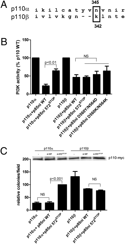
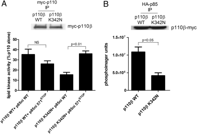
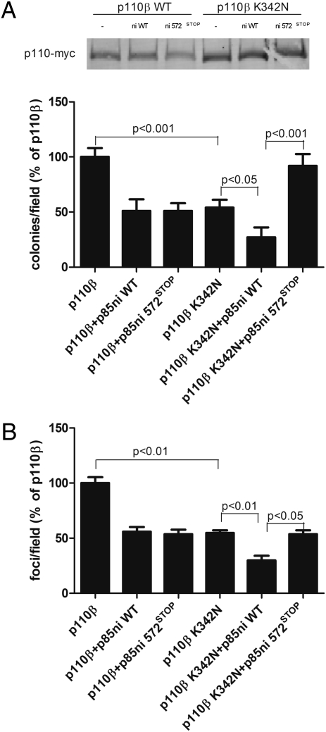
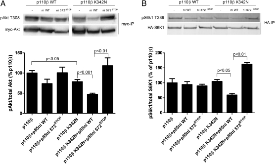
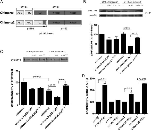
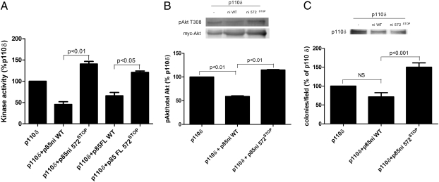
Class I PI3-kinases signal downstream of receptor tyrosine kinases and G protein-coupled receptors and have been implicated in tumorigenesis. Although the oncogenic potential of the PI3-kinase subunit p110α requires its mutational activation, other p110 isoforms can induce transformation when overexpressed in the wild-type state. In wild-type p110α, N345 in the C2 domain forms hydrogen bonds with D560 and N564 in the inter-SH2 (iSH2) domain of p85, and mutations of p110α or p85 that disrupt this interface lead to increased basal activity and transformation. Sequence analysis reveals that N345 in p110α aligns with K342 in p110β. This difference makes wild-type p110β analogous to a previously described oncogenic mutant, p110α-N345K. We now show that p110β is inhibited by p85 to a lesser extent than p110α and is not differentially inhibited by wild-type p85 versus p85 mutants that disrupt the C2-iSH2 domain interface. Similar results were seen in soft agar and focus-formation assays, where p110β was similar to p110α-N345K in transforming potential. Inhibition of p110β by p85 was enhanced by a K342N mutation in p110β, which led to decreased activity in vitro, decreased basal Akt and ribosomal protein S6 kinase (S6K1) activation, and decreased transformation in NIH 3T3 cells. Moreover, unlike wild-type p110β, p110β-K342N was differentially regulated by wild-type and mutant p85, suggesting that the inhibitory C2-iSH2 interface is functional in this mutant. This study shows that the enhanced transforming potential of p110β is the result of its decreased inhibition by p85, due to the disruption of an inhibitory C2-iSH2 domain interface.


Conflict of interest statement

The authors declare no conflict of interest.

Figures

Fig. 1.
Fig. 1.
Regulation of p110β activity and transformation potential by p85. (A) Sequence alignment of p110α and p110β showing the position of the N345 position in p110α relative to K342 in p110β. (B) Recombinant p110α or p110β, produced in insect cells, was incubated for 1 h at 4 °C without or with 1 μg of wild-type or mutant p85ni and assayed for lipid kinase activity at 22 °C as described. Data are the mean ± SEM of triplicate samples from three experiments. (C) NIH 3T3 cells were transiently transfected with p110α-myc or p110β-myc alone or with wild-type or mutant p85ni. The top panel shows p110 expression levels. The cells were plated in soft agar as described, and colonies were counted after 3 wk. Colony counts are normalized to the number produced by cells expressing p110α plus p85ni572STOP. Data are the mean ± SEM of triplicate samples from three experiments. NS, not significant.
Fig. 2.
Fig. 2.
Regulation of wild-type p110β and p110β-K342N by p85. (A) HEK 293T cells were transfected with wild-type or K342N myc-p110β. Anti-myc immunoprecipitates were incubated for 1 h at 4 °C with wild-type or mutant p85ni and were assayed for lipid kinase activity at 22 °C. Data are the mean ± SEM of triplicate samples from two experiments. The top panel shows p110 expression levels. (B) HEK 293T cells were transfected with wild-type or K342N p110β-myc and HA-tagged wild-type p85. Anti-HA immunoprecipitates were analyzed for myc-p110β levels by Western blotting (Upper) and for lipid kinase activity (Lower). Data are the mean ± SEM of triplicate samples from two experiments.
Fig. 3.
Fig. 3.
Transformation potential of wild-type p110β or p110β-K342N. (A) NIH 3T3 cells were transiently transfected with wild-type or K342N p110β-myc alone or with wild-type or mutant p85ni. The top panel shows p110 expression levels. The cells were plated in soft agar, and colonies were counted after 3 wk. Colony counts were normalized to the number produced by cells expressing p110β alone. Data are the mean ± SEM of triplicate samples from three experiments. (B) 3T3 cells were transiently transfected with wild-type or K342N p110β-myc alone or with wild-type or mutant p85ni and were left to grow to confluence for 2 wk. Foci were counted and normalized to the number produced by cells expressing p110β alone. Data are the mean ± SEM of two experiments.
Fig. 4.
Fig. 4.
Signaling downstream of wild-type or K342N p110β. (A) HEK 293T cells were transiently transfected with myc-Akt plus wild-type or K342N p110β-myc alone or with wild-type or mutant p85ni. Cells then were starved overnight, and Akt immunoprecipitates were blotted with anti-pT308-Akt antibody. Data are the mean ± SEM of three experiments. (B) HEK 293T cells were transiently transfected with HA-S6K1 plus wild-type or K342N p110β-myc, alone or with wild-type or mutant p85ni. Cells then were starved overnight, and S6K1 immunoprecipitates were blotted with anti-pT389-S6K1 antibody. Data are the mean ± SEM from two or three experiments.
Fig. 5.
Fig. 5.
Transformation potential of p110α/β chimeras. (A) Schematic representation of the structures of the two chimeric p110α/β molecules. Construction of the chimeras is described in detail in Materials and Methods. (B) HEK 293T cells were transiently transfected with myc-Akt plus myc-tagged chimera 1 or chimera 2 alone or with wild-type or mutant p85ni. Cells were starved overnight, and Akt immunoprecipitates were blotted with anti-pT308-Akt antibody. Data are the mean ± SEM of two experiments. (C) NIH 3T3 cells were transiently transfected with myc-tagged chimera 1 or chimera 2 alone or with wild-type or mutant p85ni. The top panel shows p110 expression levels. The cells were plated in soft agar, and colonies were counted after 3 wk. Colony counts were normalized to the number produced by cells expressing chimera 1 alone. (D) HEK 293T cells were transfected with myc-Akt and p85 plus p110α-myc, p110β-myc, or myc-tagged chimera 1 or chimera 2, with or without Gβγ subunits. Akt immunoprecipitates were blotted with anti-Akt and anti-pT308-Akt antibodies. Data are the mean ± SEM of three experiments.
Fig. 6.
Fig. 6.
Regulation of p110δ activity, signaling, and transformation by wild-type and mutant p85. (A) HEK 293T cells were transfected with myc-p110δ as described in Materials and Methods. Anti-myc immunoprecipitates were incubated for 1 h at 4 °C with wild-type or mutant full-length p85 and were assayed for lipid kinase activity at 22 °C. Data are the mean ± SEM of triplicate samples from two experiments. (B) HEK 293T cells were transiently transfected with myc-Akt, plus untagged p110δ alone or with wild-type or mutant p85ni. Cells then were starved overnight, and Akt immunoprecipitates were blotted with anti-pT308-Akt antibody. Data are the mean ± SEM of two experiments. (C) NIH 3T3 cells were transiently transfected with untagged p110δ alone or with wild-type or mutant p85ni. The top panel shows p110 expression levels. The cells were plated in soft agar, and colonies were counted after 3 wk. Colony counts are normalized to the number produced by cells expressing p110δ alone. Data are the mean ± SEM of triplicate samples from two experiments.

References

    1. Engelman JA, Luo J, Cantley LC. The evolution of phosphatidylinositol 3-kinases as regulators of growth and metabolism. Nat Rev Genet. 2006;7:606–619. - PubMed
    1. Yuan TL, Cantley LC. PI3K pathway alterations in cancer: Variations on a theme. Oncogene. 2008;27:5497–5510. - PMC - PubMed
    1. Geering B, Cutillas PR, Nock G, Gharbi SI, Vanhaesebroeck B. Class IA phosphoinositide 3-kinases are obligate p85-p110 heterodimers. Proc Natl Acad Sci USA. 2007;104:7809–7814. - PMC - PubMed
    1. Kurosu H, et al. Heterodimeric phosphoinositide 3-kinase consisting of p85 and p110beta is synergistically activated by the betagamma subunits of G proteins and phosphotyrosyl peptide. J Biol Chem. 1997;272:24252–24256. - PubMed
    1. Guillermet-Guibert J, et al. The p110beta isoform of phosphoinositide 3-kinase signals downstream of G protein-coupled receptors and is functionally redundant with p110gamma. Proc Natl Acad Sci USA. 2008;105:8292–8297. - PMC - PubMed

Publication types

MeSH terms

Substances