. 2010 Sep 12:16:1864-73.

Subtoxic levels hydrogen peroxide-induced production of interleukin-6 by retinal pigment epithelial cells

Affiliations

Subtoxic levels hydrogen peroxide-induced production of interleukin-6 by retinal pigment epithelial cells

Wen-Chuan Wu et al. Mol Vis. .

Abstract

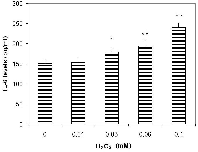
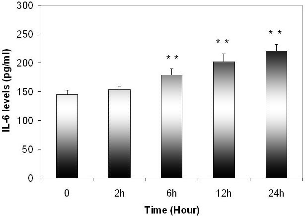
Purpose: To study the effect of subtoxic levels of hydrogen peroxide (H(2)O(2)) on the expression and release of interleukin-6 (IL-6) by cultured retinal pigment epithelial (RPE) cells and to explore the relevant signal pathways.

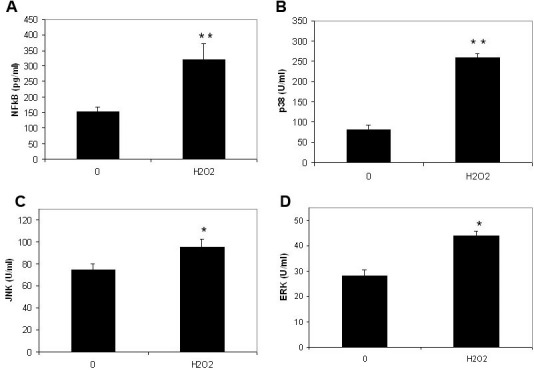
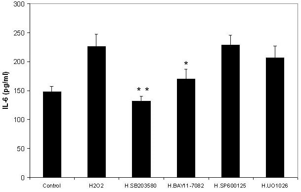
Methods: Cultured human RPE cells were stimulated with various subtoxic concentrations of H(2)O(2) for different periods. Conditioned medium and cells were collected. IL-6 in the medium and IL-6 mRNA in the collected cells were measured using an IL-6 enzyme-linked immunosorbent assay kit and reverse transcriptase polymerase chain reaction, respectively. Nuclear factor-kappaB (NF-κB) in nuclear extracts and phosphorylated p38 mitogen-activated protein kinase (MAPK), extracellular signal-regulated kinase (ERK), and c-Jun N-terminal kinases (JNK) in cells cultured with and without H(2)O(2) were measured by NF-κB and MAPK enzyme-linked immunosorbent assay kits. Inhibitors of p38 (SB203580), ERK (UO1026), JNK (SP600125), and NF-κB (BAY11-7082) were added to the cultures before the addition of H(2)O(2) to test their effects(.)

Results: Subtoxic levels of H(2)O(2) (100 µM and less) increased the IL-6 mRNA level and the release of IL-6 protein by the cultured human RPE cells in a dose- and time-dependent manner. This was accompanied by an increase of NF-κB in nuclear extracts and phosphorylated p38 MAPK, ERK, and JNK in cell lysates, particularly in the p38 and NF-κB. The NF-κB inhibitor decreased the H(2)O(2)-induced expression of IL-6. The p38 inhibitor, but not the ERK or JNK inhibitor, completely abolished H(2)O(2)-induced expression of IL-6 by RPE cells. The p38 inhibitor also abolished the increase of NF-κB in nuclear extracts in cells treated with H(2)O(2).

Conclusions: H(2)O(2) stimulated the production of IL-6, a key factor in the modulation of immune responses, inflammatory processes, and the occurrence of autoimmune diseases, which recently has been documented to be increased in age-related macular degeneration (AMD). This may be a molecular linkage for the oxidative stress and inflammatory/autoimmune reactions in AMD and may provide a novel target for the treatment of AMD.


Figures

Figure 1
Figure 1
The dose-dependent effects of subtoxic levels of hydrogen peroxide on release of interleukin-6 by retinal pigment epithelium cells. Retinal pigment epithelium (RPE) cells (ARPE-19) were plated into 24-well plates. After 24 h incubation, hydrogen peroxide (H2O2, at 0, 10, 30, 60, and 100 µM) was added to the culture and incubated for 24 h. Conditioned culture medium was collected and the amount of interleukin-6 (IL-6) protein in the conditioned medium was determined using the human IL-6 Quantikine enzyme-linked immunosorbent assay (ELISA) kit. IL-6 levels in conditioned culture medium were expressed as pg/ml (mean±standard deviation in triplicate tests). *0.01< p<0.05, **p<0.01, compared with the controls (cells cultured without H2O2).
Figure 2
Figure 2
The time-dependent effects of subtoxic levels of hydrogen peroxide on release of interleukin-6 by retinal pigment epithelium cells. Retinal pigment epithelium (RPE) cells (ARPE-19) were plated into 24-well plates. After 24 h incubation, 100 µM hydrogen peroxide (H2O2) was added to the culture and incubated for 2, 6, 12, and 24 h. Conditioned culture medium was collected and the amount of interleukin-6 (IL-6) protein in the conditioned medium was determined by using the human IL-6 Quantikine enzyme-linked immunosorbent assay (ELISA) kit. IL-6 levels in conditioned culture medium were expressed as pg/ml (mean±standard deviation in triplicate tests). *0.01< p<0.05, **p<0.01, compared with the controls (cells cultured without H2O2).
Figure 3
Figure 3
The effects of subtoxic levels of hydrogen peroxide on nuclear factor factor-kappaB in nuclear extracts and phosphorylated extracellular signal-regulated kinases, c-Jun N-terminal kinase and p38 mitogen-activated protein kinase. Retinal pigment epithelium (RPE) cells (ARPE-19) were plated into 24-well plates. After 24 h incubation, 100 µm hydrogen peroxide (H2O2) was added to the medium. Cells were collected 30 min later. The nuclear factor factor-kappaB (NF-κB) levels in nuclear extracts (A) and phosphorylated p38 mitogen-activated protein kinase (p38; B), c-Jun N-terminal kinase (JNK; C) and extracellular signal-regulated kinases (ERK; D) in cell lysates were measured using the relevant NF-κB enzyme-linked immunosorbent assay (ELISA) kit and phosphorylated MAPK ELISA kits (Biosource). The levels of NF-κB in nuclear extracts were expressed as pg/ml and phosphorylated p38, ERK and JNK in cell lysates were expressed as U/ml (mean±standard deviation in triplicate tests). *0.01< p<0.05, **p<0.01, compared with the controls (cells cultured without H2O2).
Figure 4
Figure 4
Effects of nuclear factor factor-kappaB and mitogen-activated protein kinase inhibitors on the hydrogen peroxide-induced production of interleukin-6 by retinal pigment epithelium cells. Retinal pigment epithelium (RPE) cells (ARPE-19) were plated into 24-well plates. After 24 h incubation, various mitogen-activated protein kinase (MAPK) and nuclear factor-kappaB (NF-κB) inhibitors were added to the medium separately, including BAY11–7082 (NF-κB inhibitor), UO1026 (extracellular signal-regulated kinases inhibitor), SP600125 (c-Jun N-terminal kinase inhibitor) and SB203580 (p38 MAPK inhibitor, all from Calbiochem) at a final concentration of 10 µM with the exception of BAY11–7082 (5 µM). After 30 min, 100 µM hydrogen peroxide (H2O2) was added to the medium. Cells cultured without H2O2 were used as negative controls. Cells cultured with H2O2 but without inhibitors were used as positive controls. After 24 h incubation, the culture medium was collected and the IL-6 levels were measured with the human IL-6 Quantikine ELISA kit and expressed as pg/ml (mean±standard deviation in triplicate tests). *0.01< p<0.05, **p<0.01, compared with the positive controls (cells cultured with H2O2 but without inhibitors).

Similar articles

Cited by

References

    1. Rein DB, Wittenborn JS, Zhang X, Honeycutt AA, Lesesne SB, Saaddine J, Vision Health Cost-Effectiveness Study Group. Forecasting age-related macular degeneration through the year 2050: the potential impact of new treatments. Arch Ophthalmol. 2009;127:533–40. - PubMed
    1. Hogg R, Chakravarthy U. AMD and micronutrient antioxidants. Curr Eye Res. 2004;29:387–401. - PubMed
    1. Winkler BS, Boulton ME, Gottsch JD, Sternberg P. Oxidative damage and age-related macular degeneration. Mol Vis. 1999;5:32. - PMC - PubMed
    1. Cai J, Nelson KC, Wu M, Sternberg P, Jr, Jones DP. Oxidative Damage and protection of the RPE. Prog Retin Eye Res. 2000;19:205–21. - PubMed
    1. Zarbin MA. Current concepts in the pathogenesis of age-related macular degeneration. Arch Ophthalmol. 2004;122:598–614. - PubMed

Publication types

MeSH terms

LinkOut - more resources