

Molecular analysis of cataract families in India: new mutations in the CRYBB2 and GJA3 genes and rare polymorphisms
- PMID: 21031021
- PMCID: PMC2956670
Molecular analysis of cataract families in India: new mutations in the CRYBB2 and GJA3 genes and rare polymorphisms
Abstract
Purpose: The aim of the study was to resolve the genetic etiology in families having inherited cataracts.
Methods: Families afflicted with congenital/childhood cataracts were registered in Chennai and Orissa (India). Blood samples were collected from the probands and available family members. Selected functional candidate genes were amplified by polymerase chain reaction (PCR) and characterized by direct sequencing. Putative mutations were confirmed in healthy controls.
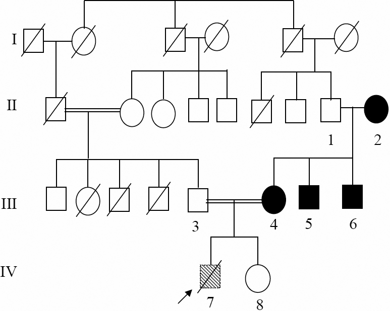
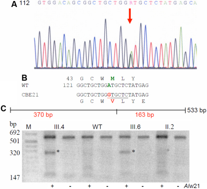
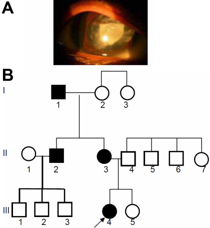
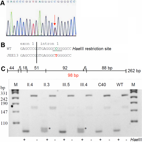
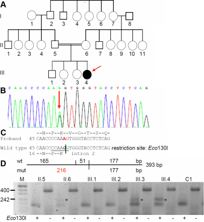
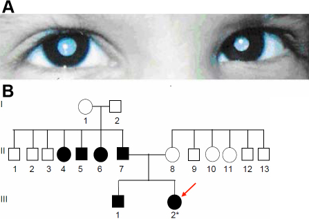
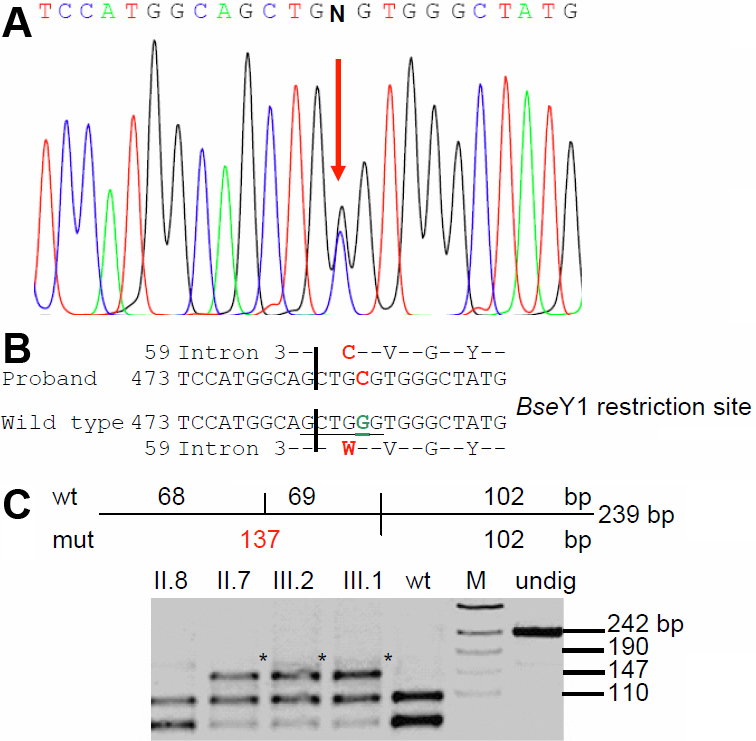
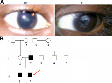
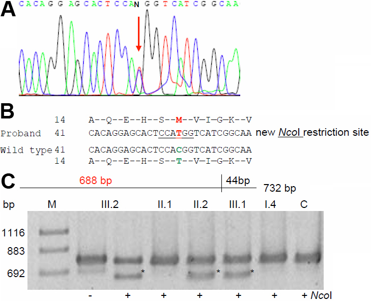
Results: We observed interesting new polymorphisms of ethnic specificity, some of frequent nature, such as a 3-bp deletion in intron 3 of CRYBB2 (encoding βB2-crystallin) and IVS1+9 c>t variation in HSF4 (encoding heat-shock factor 4). Some rare single nucleotide polymorphisms (SNPs) co-segregate with the respective phenotype such as IVS3+120c>a of CRYBB2, while M44V of CRYGD (encoding γD-crystallin), although found in association with blue dot opacity was seen in a few healthy controls too. We identified two new mutations co-segregating along with the respective cataract phenotype within the families that were not seen in healthy controls from India or Germany. These include two missense mutations; one in GJA3 (encoding gap junction protein α3, which is also referred to as connexin 46); the mutation affects codon 19 (T19M), and the corresponding phenotype is a posterior-polar cataract. The other missense mutation affects CRYBB2 (W59C; total cataract). Additionally, a cDNA variation (G54A) identified in a zonular cataract affects a highly conserved splice site of CRYBB2. This mutation, however, showed reduced penetrance in the family, which might be explained by different molecular consequences in the affected family members: nonsense-mediated decay of the mutated mRNA might have no clinical phenotype in heterozygotes, whereas the translation of the mutated mRNA is predicted to lead to a small hybrid protein (consisting of 16 amino acids of the βB2-crystallin and 18 new amino-acids), which might have a dominant-negative function in the lens.
Conclusions: This report identifies in families with childhood cataract some new alleles, which may be considered as causative for cataracts. Furthermore, we report some geographically restricted rare polymorphic sites, whose significance might be considered in some context as modifiers or alleles in sensitizing ocular lens toward cataractogenesis.
Figures
Similar articles
-
Mutation analysis of congenital cataracts in Indian families: identification of SNPS and a new causative allele in CRYBB2 gene.Invest Ophthalmol Vis Sci. 2004 Oct;45(10):3599-607. doi: 10.1167/iovs.04-0207. Invest Ophthalmol Vis Sci. 2004. PMID: 15452067
-
A novel human CRYGD mutation in a juvenile autosomal dominant cataract.Mol Vis. 2010 May 22;16:887-96. Mol Vis. 2010. PMID: 20508808 Free PMC article.
-
Comprehensive mutational screening in a cohort of Danish families with hereditary congenital cataract.Invest Ophthalmol Vis Sci. 2009 Jul;50(7):3291-303. doi: 10.1167/iovs.08-3149. Epub 2009 Jan 31. Invest Ophthalmol Vis Sci. 2009. PMID: 19182255
-
Genetics of crystallins: cataract and beyond.Exp Eye Res. 2009 Feb;88(2):173-89. doi: 10.1016/j.exer.2008.10.011. Epub 2008 Nov 1. Exp Eye Res. 2009. PMID: 19007775 Review.
-
Identification of a novel missense mutation of MAF in a Japanese family with congenital cataract by whole exome sequencing: a clinical report and review of literature.Am J Med Genet A. 2014 May;164A(5):1272-6. doi: 10.1002/ajmg.a.36433. Epub 2014 Mar 24. Am J Med Genet A. 2014. PMID: 24664492 Review.
Cited by
-
Whole Exome Sequencing Reveals a Mutation in CRYBB2 in a Large Mexican Family with Autosomal Dominant Pulverulent Cataract.Mol Syndromol. 2016 May;7(2):87-92. doi: 10.1159/000445669. Epub 2016 Apr 14. Mol Syndromol. 2016. PMID: 27385965 Free PMC article.
-
The connexin46 mutant, Cx46T19M, causes loss of gap junction function and alters hemi-channel gating.J Membr Biol. 2015 Feb;248(1):145-55. doi: 10.1007/s00232-014-9752-y. Epub 2014 Nov 18. J Membr Biol. 2015. PMID: 25404239 Free PMC article.
-
High-Throughput Genetic Screening of 51 Pediatric Cataract Genes Identifies Causative Mutations in Inherited Pediatric Cataract in South Eastern Australia.G3 (Bethesda). 2017 Oct 5;7(10):3257-3268. doi: 10.1534/g3.117.300109. G3 (Bethesda). 2017. PMID: 28839118 Free PMC article.
-
Sporadic and Familial Congenital Cataracts: Mutational Spectrum and New Diagnoses Using Next-Generation Sequencing.Hum Mutat. 2016 Apr;37(4):371-84. doi: 10.1002/humu.22948. Epub 2016 Jan 14. Hum Mutat. 2016. PMID: 26694549 Free PMC article.
-
Novel CRYGC Mutation in Conserved Ultraviolet-Protective Tryptophan (p.Trp131Arg) Is Linked to Autosomal Dominant Congenital Cataract.Int J Mol Sci. 2023 Nov 22;24(23):16594. doi: 10.3390/ijms242316594. Int J Mol Sci. 2023. PMID: 38068917 Free PMC article.
References
-
- Francis PJ, Moore AT. Genetics of childhood cataract. Curr Opin Ophthalmol. 2004;15:10–5. - PubMed
-
- Dandona L, Williams JD, Williams BC, Rao GN. Population-based assessment of childhood blindness in Southern India. Arch Ophthalmol. 1998;116:545–6. - PubMed
-
- Graw J. The genetic and molecular basis of congenital eye defects. Nat Rev Genet. 2003;4:876–88. - PubMed
-
- Graw J. Mouse Models for Cataracts. J Genet. 2009;88:469–86. - PubMed
-
- Graw J. Genetics of crystallins: Cataract and beyond. Exp Eye Res. 2009;88:173–89. - PubMed
Publication types
MeSH terms
Substances
LinkOut - more resources
Full Text Sources
Medical
Miscellaneous