. 2011:692:159-71.
doi: 10.1007/978-1-60761-971-0_12.

Defining the structure and function of acyl-homoserine lactone autoinducers

Affiliations

Defining the structure and function of acyl-homoserine lactone autoinducers

Mair E A Churchill et al. Methods Mol Biol. 2011.

Abstract

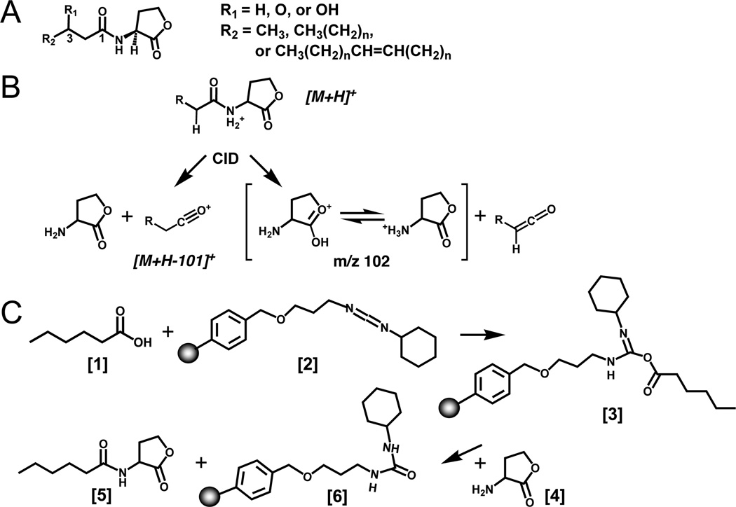
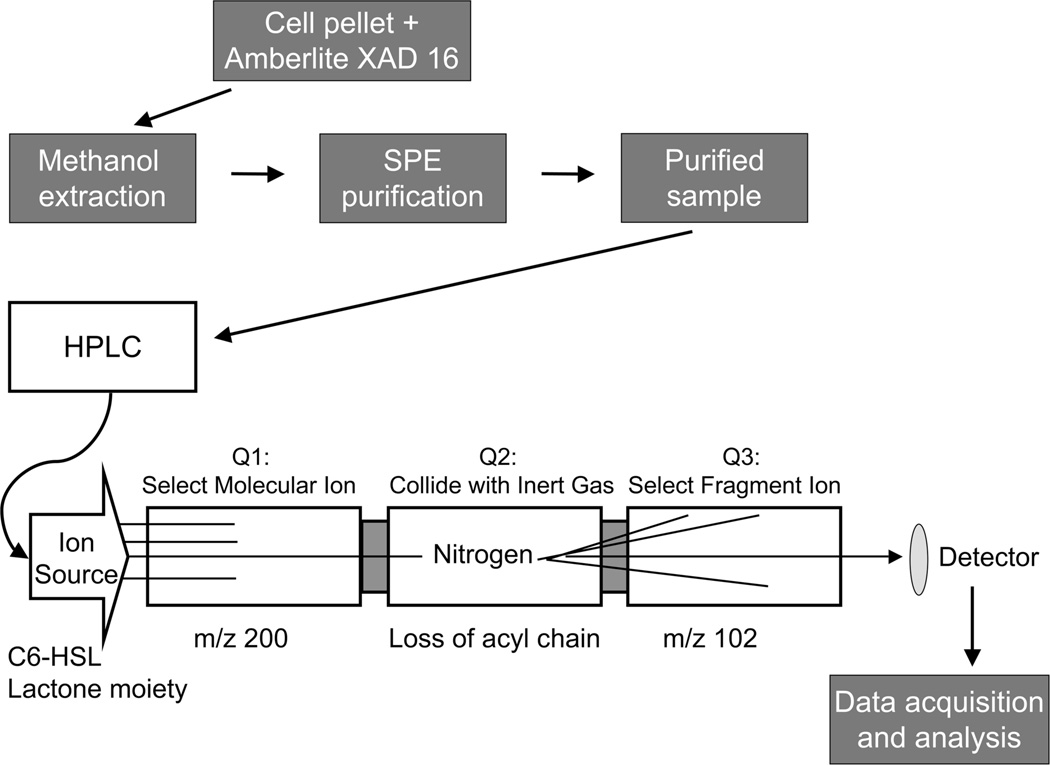
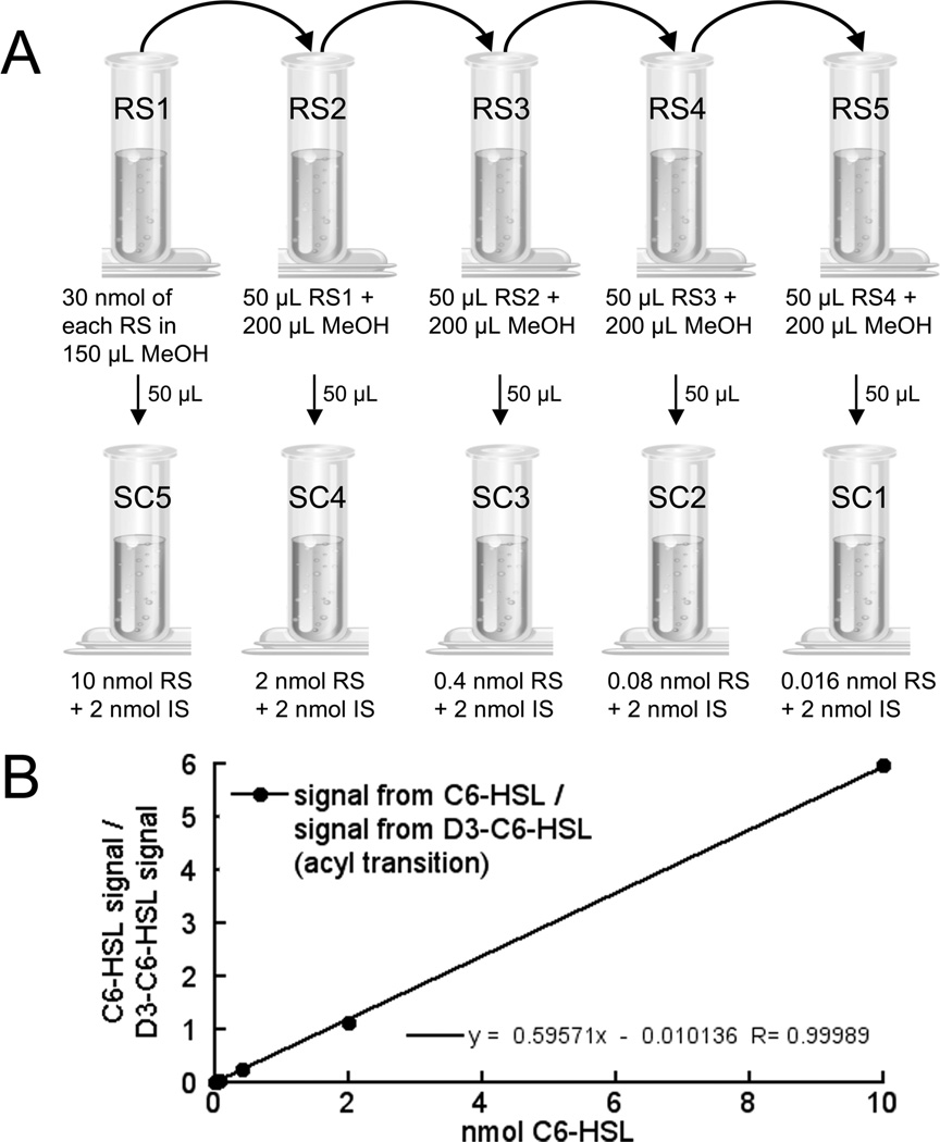
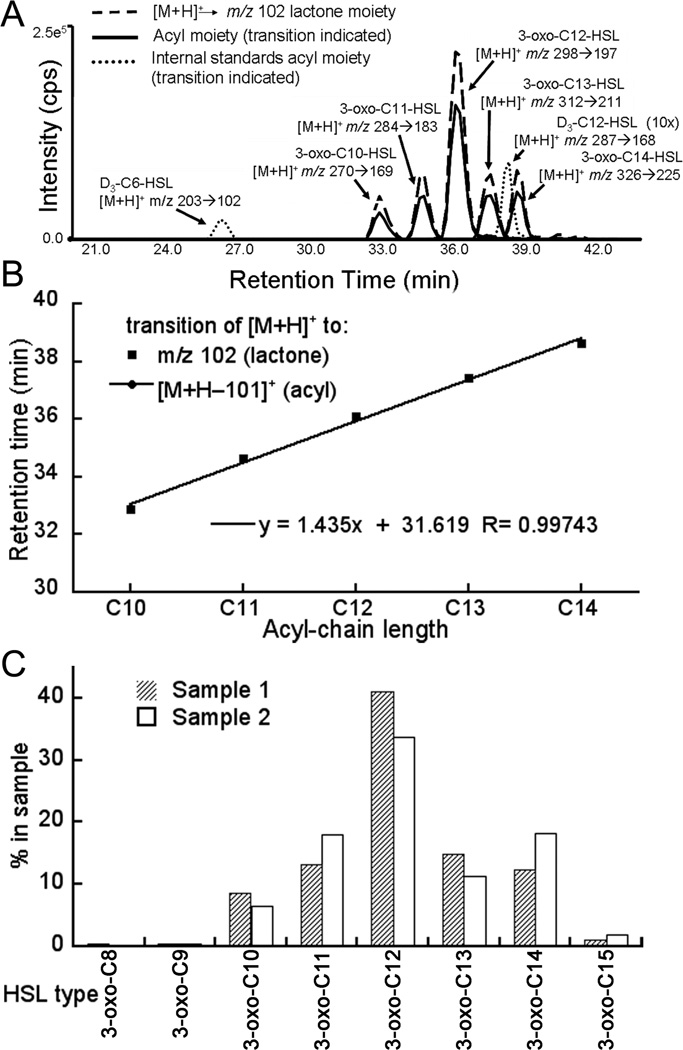
Quorum sensing plays a central role in regulating many community-derived symbiotic and pathogenic relationships of bacteria, and as such has attracted much attention in recent years. Acyl-homoserine lactones (AHLs) are important signaling molecules in the quorum sensing gene-regulatory processes found in numerous gram-negative species of bacteria that interact with eukaryotic organisms. AHLs are produced by acyl-homoserine lactone synthases. Bacteria can have multiple genes for AHL synthase enzymes, and such species are likely to produce several different types of AHLs. Determination of the types and the relative amounts of AHLs produced by AHL synthases in bacteria under varied conditions provides important insights into the mechanism of AHL synthase function and the regulation of transcriptional cascades initiated by quorum sensing signaling. This chapter describes a mass spectrometry method for determining the types and relative amounts of AHLs present in a sample.


Figures

Figure 1
Figure 1
Scheme of AHL structure, mass spectrometry, and chemical synthesis. (A) Structure of AHLs. AHLs vary by substitution at the C3 position (R1) and the length and unsaturation of the acyl chain (R2). HSL refers to D/L-homoserine lactone; AHL refers to N-acyl-D/L-homoserine lactone, with any acyl chain length (indicated by the number of carbons CN) or degree of substitution, and the extent of deuterium labeling indicated by Dn. (B) Mass spectrometry scheme proposed for collisionally induced decomposition of AHLs into the two major fragment ions observed for the 3-oxo-HSLs (lactone and acyl). (C) Carbodiimide based chemical synthesis of D3-C6-HSL. The fatty acid [1] reacts with the resin bound carbodiimide reagent [2] to give an acylating intermediate [3]. Next, nucleophilic attack by the HSLhydrobromide amine [4] on the acylating intermediate releases the amide product AHL [5]. A urea-like byproduct remains coupled to the resin-bound carbodiimide [6]. Any excess or unreacted fatty acid was removed during filtration because it is left in the form of the acylating intermediate bound to the resin [6]. The final AHL product [5] was obtained by filtration of the reaction mixture and solid-phase extraction using a normal-phase SPE.
Figure 2
Figure 2
Flowchart of the methods described in this chapter. The cell cultures were grown in the presence of Amberlite XAD 16, which adsorbs AHLs. In order to extract the AHLs from the resin and cell debris, the harvested pellet from the bacterial culture and Amberlite XAD 16 was re-suspended in methanol and left overnight. The methanol solution was filtered and centrifuged, and then subjected to solid phase extraction to remove any remaining impurities and to concentrate the AHLs. The purified sample was then injected onto the reversed phase HPLC column to separate the AHLs based on their lipophilicity and introduce them into the mass spectrometer. In the multiple reaction monitoring (MRM) experiment, the sample was passed through the ion source into Quadrupole1 (Q1), then the selected ions proceeded into Quadrupole 2 (Q2) where they collide with inert gas (nitrogen). After fragmentation, selected ions move into Quadrupole 3 (Q3) and are visualized by the detector. Data were processed by applying standard isotope dilution curves to the analytical data (as in Figures 3 and 4).
Figure 3
Figure 3
Preparation of a standard curve for mass spectrometry of AHLs. (A) Step-by-step preparation of internal standard and reference standard mixtures for mass spectrometry. First a series of serial dilutions (1:5) are prepared from a stock solution (vial 1A) containing 30 nmol each of reference standard (RS) in 150 µL methanol (Table 3). Here the four reference standards being used are 3-oxo-C6-HSL, 3-oxo-C12-HSL, C6-HSL, and C12-HSL. The samples in vials 2A, 3A, 4A, and 5A are then made by serial dilution of 50 µL from the preceding vial into 200 µL of methanol. To produce the standard curve samples (SC1–5), 50 µL of each samples 1A–5A is transferred to a new vial and a fixed amount of the stable isotope labeled internal standard (IS) is added to each vial, as well as an empty control vial (SC0, not shown in the figure). Here the IS was D3-C6-HSL and 2 nmol was added to each of the SC vials. The samples are then dried down before use. (B) Standard curve of C6-HSL. The ratio of the integrated ion transition peak area of a particular transition for the RS (signal of C6-HSL [M+H]+ –> [M+H-101]+) to the integrated ion transition peak area of the same transition of the IS (signal of D3-C6-HSL [M+H]+ –> [M+H-101]+) was plotted and fitted with a linear equation (black line). This standard curve was then applied to analytical data.
Figure 4
Figure 4
Example of AHL analysis of AHLs produced by LasI expressed in E. coli. (A) Total ion current observed in the MRM analysis of reversed phase chromatographic separation of AHLs. AHLs were examined using the MRM mode that was monitoring the transitions for precursor [M+H]+ –> m/z 102 (shown as a dashed line) and the transition from [M+H]+ –> [M+H-101]+ (shown as solid lines), and the IS peaks (dotted lines). The identified AHLs are labeled. In this experiment, it is clear that the integrated peak areas for the ion transitions of 3-oxo-AHLs are of nearly equally size (approximately 40:60) for the two transitions (to the acyl and lactone moieties, repectively). (B) Retention times of the transitions observed for the lactone and acyl moieties plotted versus the number of carbon atoms in the acyl chain of each AHL. The plot shows that all the AHLs with the same substitution lie on a straight line, which coincides with a line that was generated by using the reference standards. Deviation from the line of the reference standards indicates that the compound, which has been named in the data analysis is likely to be incorrect. (C) Bar graph representation of the AHLs purified from two independent experiments of LasI expressed in E. coli, with two slightly different growth and extraction conditions. Sample 1 and sample 2 differ in number of hours that LasI was grown as well as the addition of Amberlite XAD 16. In sample 1, the Amberlite XAD 16 resin was added immediately after induction and the culture was grown for only eight hours, whereas for sample 2 the Amberlite XAD 16 resin was added right before inoculation and the culture was grown for 24 hours. The height of the bars in the graph represents the percentage of each AHL and not the total amounts of AHL in the sample.

References

    1. Fuqua C, Eberhard A. Signal generation in autoinduction systems: synthesis of acylated homoserine lactones by LuxI-type proteins. In: Dunny G, Winans SC, editors. Cell-cell signaling in bacteria. Washington, D.C.: ASM Press; 1999. pp. 211–230.
    1. Pearson JP, Passador L, Iglewski BH, Greenberg EP. A second N-acylhomoserine lactone signal produced by Pseudomonas aeruginosa. Proc Natl Acad Sci U S A. 1995;92:1490–1494. - PMC - PubMed
    1. Churchill MEA, Herman JP. Acyl-homoserinelactone Biosynthesis: Structure and Mechanism." Chapter 17. In: Winans Stephen, Bassler Bonnie., editors. Chemical Communication Among Bacteria. Washington, D.C.: ASM Press; 2008.
    1. Shaw PD, Ping G, Daly SL, Cha C, Cronan JE, Rinehart KL, Farrand SK. Detecting and characterizing N-acyl-homoserine lactone signal molecules by thin-layer chromatography. Proc. Natl. Acad. Sci U.S.A. 1997;94:6036–6041. - PMC - PubMed
    1. Gould TA, Herman JP, Krank J, Murphy RC, Churchill MEA. Specificity of acyl-homoserine lactone syntheses examined by mass spectrometry. J Bacteriol. 2006;188:773–783. - PMC - PubMed

Publication types

MeSH terms

LinkOut - more resources