. 2011 Jan;20(1):42-50.
doi: 10.1002/pro.534.

Cryoprotective mechanism of a small intrinsically disordered dehydrin protein

Affiliations

Cryoprotective mechanism of a small intrinsically disordered dehydrin protein

Stephanie Hughes et al. Protein Sci. 2011 Jan.

Abstract

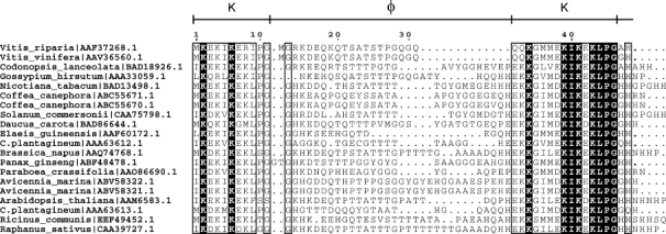
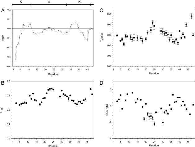
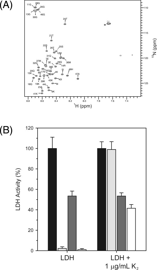
Dehydration proteins (Dehydrins) are expressed during dehydration stress in plants and are thought to protect plant proteins and membranes from the loss of water during drought and at cold temperatures. Several different dehydrins have been shown to protect lactate dehydrogenase (LDH) from damage from being frozen and thawed. We show here that a 48 residue K₂ dehydrin from Vitis riparia protects LDH more effectively than bovine serum albumin, a protein with known cryoprotective function. Light scattering and 8-anilino-1-naphthalene sulfonate fluorescence experiments show that dehydrins prevent aggregation and unfolding of the enzyme. The cryoprotective effects of LDH are reduced by the addition of salt, suggesting that the positively charged K-segments are attracted to a negatively charged surface but this does not result in binding. Overall K₂ is an intrinsically disordered protein; nuclear magnetic resonance relaxation experiments indicate that the two-terminal, Lys-rich K-segments show a weak propensity for α-helicity and are flexible, and that the central, polar rich phi-segment has no secondary structure preference and is highly flexible. We propose that the phi-segments in dehydrins are important for maintaining the disordered structure so that the protein can act as a molecular shield to prevent partially denatured proteins from interacting with one another, whereas the K-segments may help to localize the dehydrin near the enzyme surface.


Figures

Figure 1
Figure 1
Alignment of K-φ-K sequences. A ClustalW generated alignment of 20 K2 regions with the highest sequence identity to Vitis riparia K2. Conserved regions are shown bound by a rectangle, where a white letter on a black background indicates a residue that is conserved in all sequences, and a black letter indicates residues that are partially conserved and/or chemically similar. The sequence numbering for Vitis riparia K2 and the segment names are shown at the top of the alignment. The organism name and protein NCBI accession number are indicated on the left. The figure was generated using ESPript.
Figure 2
Figure 2
Structure and dynamics of K2 in solution. The arrangement of the K- and φ-segments is shown at the top of the figure. A: The SSP scores of K2 were calculated using Hα, Cα, and Cβ chemical shifts and are plotted on a per residue basis. B: T1, (C) T2, and (D) 15N-NOE relaxation data of K2. Error bars represent the error in fitting the relaxation decay curves as described in CCPNMR.
Figure 3
Figure 3
Cryoprotection of LDH. The protection of LDH activity after freezing and thawing of the enzyme was examined in the presence and absence of additives (K2, YSK2, BSA, or lysozyme) over a range of concentrations. The activity of unfrozen LDH is defined as 100% activity. K2, solid circle; YSK2, open circle; BSA, open square; lysozyme, solid triangle. Error bars represent the standard deviation of n = 6 (K2, BSA, lysozyme) or n = 3 (YSK2). The lines represent fits to the sigmoidal equation: % LDH activity =formula image
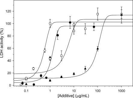
, where x is the concentration of the additive.
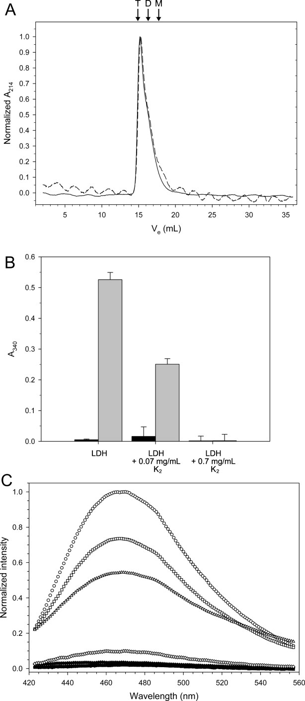
Figure 4
Figure 4
Effect of freezing and freeze-protection on the structure of LDH. A: Gel filtration of 200 μg of LDH before (solid line) and after (dashed line) being frozen and thawed. The arrows and labels indicate the predicted elution volume of the various oligomeric states: T, tetramer; D, dimer; M, monomer. Ve represents the elution volume of the sample. B: Light scatter measured at 340 nm. The bar graph represents data from n = 6 measurements, and the error bars show their standard deviation. The sample conditions are listed below each entry, where black represents unfrozen samples and gray represents frozen and thawed samples. C: Emission scans of ANS fluorescence of 1 μM LDH in the presence and the absence of K2. Samples were scanned before (solid symbols) and after (open symbols) freeze-thaw treatment. Symbols: LDH alone, circle; LDH with 0.028 mg/mL K2, square; 0.28 mg/mL K2, triangle; 0.65 mg/mL K2, diamond.
Figure 5
Figure 5
K2 interaction with LDH. (A) 15N-HSQC of 0.1 mM of K2 collected in the absence (black contours) or the presence (gray contours) of 0.3 mM LDH. The peak labels show the residue number and single letter amino acid code of K2. B: Cryoprotection of LDH by K2 is mediated by weak electrostatic interactions. The bar graph shows the percent LDH activity of samples assayed in the presence and the absence of salt with and without 1 μg/mL K2. The data represent n = 6 measurements with the error bar corresponding to the standard deviation. Unfrozen samples with no NaCl, black bars; frozen samples with no NaCl, light gray; unfrozen samples with 1M NaCl, dark gray; frozen samples with 1M NaCl, white.

Similar articles

Cited by

References

    1. Close TJ. Dehydrins: a commonality in the response of plants to dehydration and low temperature. Physiol Plant. 1997;100:291–296.
    1. Rorat T. Plant dehydrins—tissue location, structure and function. Cell Mol Biol Lett. 2006;11:536–556. - PMC - PubMed
    1. Kosova K, Vitamvas P, Prasil IT. The role of dehydrins in plant response to cold. Biol Plant. 2007;51:601–617.
    1. Allagulova CR, Gimalov FR, Shakirova FM, Vakhitov VA. The plant dehydrins: structure and putative functions. Biochemistry. 2003;68:945–951. - PubMed
    1. Goyal K, Walton LJ, Tunnacliffe A. LEA proteins prevent protein aggregation due to water stress. Biochem J. 2005;388:151–157. - PMC - PubMed

Publication types

LinkOut - more resources