

Combined off-resonance imaging and T2 relaxation in the rotating frame for positive contrast MR imaging of infection in a murine burn model
- PMID: 21031524
- PMCID: PMC2975424
- DOI: 10.1002/jmri.22349
Combined off-resonance imaging and T2 relaxation in the rotating frame for positive contrast MR imaging of infection in a murine burn model
Erratum in
- J Magn Reson Imaging. 2011 Jan;33(1):256. Righi, Valeria [added]
Abstract
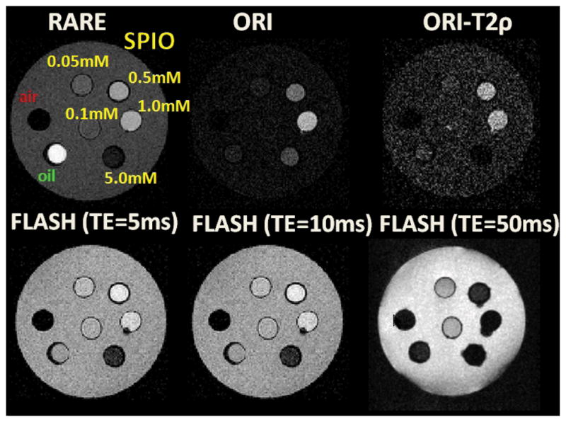
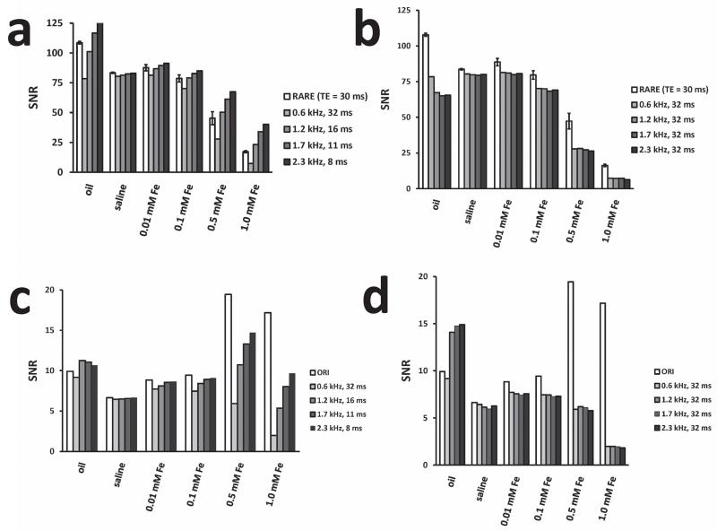
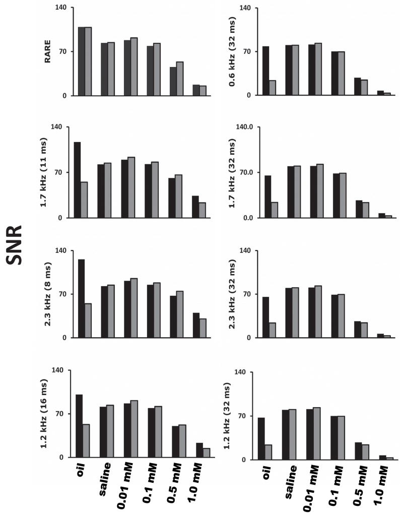
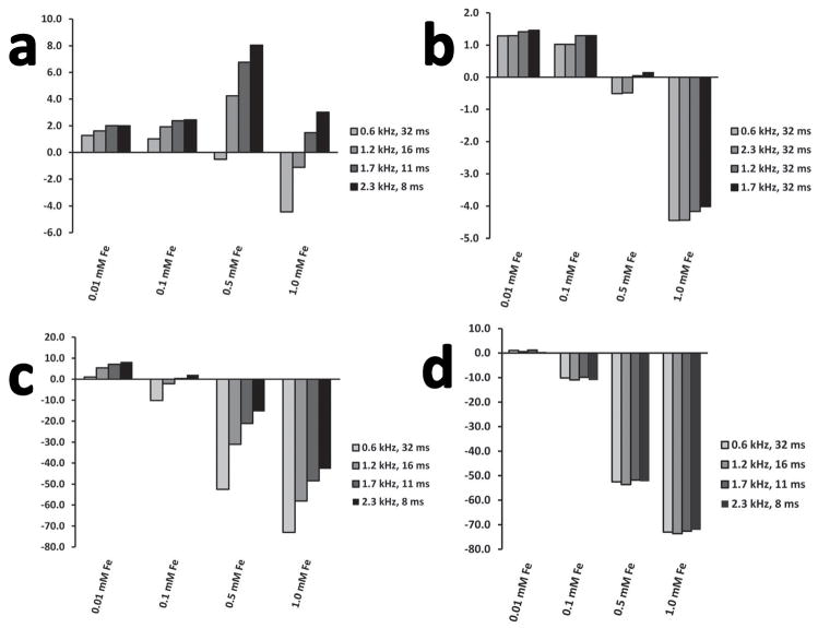
Purpose: To develop novel magnetic resonance (MR) imaging methods to monitor accumulation of macrophages in inflammation and infection. Positive-contrast MR imaging provides an alternative to negative-contrast MRI, exploiting the chemical shift induced by ultra-small superparamagnetic iron-oxide (USPIO) nanoparticles to nearby water molecules. We introduce a novel combination of off-resonance (ORI) positive-contrast MRI and T(2ρ) relaxation in the rotating frame (ORI-T(2ρ)) for positive-contrast MR imaging of USPIO.
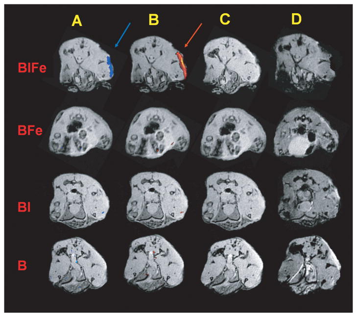
Materials and methods: We tested ORI-T(2ρ) in phantoms and imaged in vivo the accumulation of USPIO-labeled macrophages at the infection site in a mouse model of burn trauma and infection with Pseudomonas aeruginosa (PA). PA infection is clinically important. The USPIO nanoparticles were injected directly in the animals in solution, and macrophage labeling occurred in vivo in the animal model.
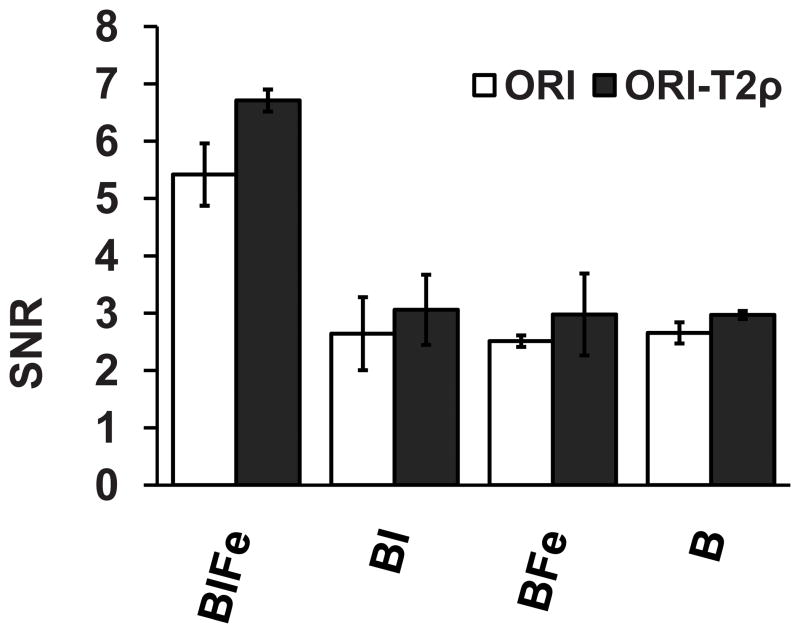
Results: We observed a significant difference between ORI-T(2ρ) and ORI, which leads us to suggest that ORI-T(2ρ) is more sensitive in detecting USPIO signal. To this end, the ORI-T(2ρ) positive contrast method may prove to be of higher utility in future research.
Conclusion: Our results may have direct implications in the longitudinal monitoring of infection, and open perspectives for testing novel anti-infective compounds.
© 2010 Wiley-Liss, Inc.
Figures

Similar articles
-
Imaging of macrophages in soft-tissue infection in rats: relationship between ultrasmall superparamagnetic iron oxide dose and MR signal characteristics.Radiology. 2005 Mar;234(3):765-75. doi: 10.1148/radiol.2343031172. Epub 2005 Jan 21. Radiology. 2005. PMID: 15665219
-
Detection of synovial macrophages in an experimental rabbit model of antigen-induced arthritis: ultrasmall superparamagnetic iron oxide-enhanced MR imaging.Radiology. 2004 Oct;233(1):149-57. doi: 10.1148/radiol.2331031402. Epub 2004 Aug 27. Radiology. 2004. PMID: 15333767
-
Septic arthritis: monitoring with USPIO-enhanced macrophage MR imaging.Radiology. 2011 Mar;258(3):722-8. doi: 10.1148/radiol.10101272. Radiology. 2011. PMID: 21339348
-
Macrophage imaging in central nervous system and in carotid atherosclerotic plaque using ultrasmall superparamagnetic iron oxide in magnetic resonance imaging.Invest Radiol. 2004 Oct;39(10):619-25. doi: 10.1097/01.rli.0000135980.08491.33. Invest Radiol. 2004. PMID: 15377941 Review.
-
Iron-based superparamagnetic nanoparticle contrast agents for MRI of infection and inflammation.AJR Am J Roentgenol. 2015 Mar;204(3):W302-13. doi: 10.2214/AJR.14.12733. AJR Am J Roentgenol. 2015. PMID: 25714316 Free PMC article. Review.
Cited by
-
T1ρ Dispersion in Articular Cartilage: Relationship to Material Properties and Macromolecular Content.Cartilage. 2015 Apr;6(2):113-22. doi: 10.1177/1947603515569529. Cartilage. 2015. PMID: 26069714 Free PMC article.
-
MRI confirms loss of blood-brain barrier integrity in a mouse model of disseminated candidiasis.NMR Biomed. 2013 Sep;26(9):1125-34. doi: 10.1002/nbm.2926. Epub 2013 Apr 22. NMR Biomed. 2013. PMID: 23606437 Free PMC article.
-
Magnetization transfer contrast MRI in GFP‑tagged live bacteria.Mol Med Rep. 2019 Jan;19(1):617-621. doi: 10.3892/mmr.2018.9669. Epub 2018 Nov 19. Mol Med Rep. 2019. PMID: 30483743 Free PMC article.
-
Tracking of Labelled Stem Cells Using Molecular MR Imaging in a Mouse Burn Model in Vivo as an Approach to Regenerative Medicine.Adv J Mol Imaging. 2021 Jan;11(1):1-15. doi: 10.4236/ami.2021.111001. Adv J Mol Imaging. 2021. PMID: 33996249 Free PMC article.
-
Imaging C-Fos Gene Expression in Burns Using Lipid Coated Spion Nanoparticles.Adv J Mol Imaging. 2012 Oct;2(4):31-37. doi: 10.4236/ami.2012.24005. Adv J Mol Imaging. 2012. PMID: 24995147 Free PMC article.
References
-
- Magnitsky S, Walton RM, Wolfe JH, Poptani H. Magnetic resonance imaging as a tool for monitoring stem cell migration. Neurodegener Dis. 2007;4(4):314–321. - PubMed
-
- Williams JB, Ye Q, Hitchens TK, Kaufman CL, Ho C. MRI detection of macrophages labeled using micrometer-sized iron oxide particles. J Magn Reson Imaging. 2007;25(6):1210–1218. - PubMed
-
- Kaim AH, Jundt G, Wischer T, et al. Functional-morphologic MR imaging with ultrasmall superparamagnetic particles of iron oxide in acute and chronic soft-tissue infection: study in rats. Radiology. 2003;227(1):169–174. - PubMed
-
- Kaim AH, Wischer T, O'Reilly T, et al. MR imaging with ultrasmall superparamagnetic iron oxide particles in experimental soft-tissue infections in rats. Radiology. 2002;225(3):808–814. - PubMed
Publication types
MeSH terms
Substances
Grants and funding
LinkOut - more resources
Full Text Sources
Other Literature Sources
Medical
