. 2010 Dec 15;518(24):4938-62.
doi: 10.1002/cne.22499.

Alternative splicing of neuroligin and its protein distribution in the outer plexiform layer of the chicken retina

Affiliations

Alternative splicing of neuroligin and its protein distribution in the outer plexiform layer of the chicken retina

Karl J Wahlin et al. J Comp Neurol. .

Abstract

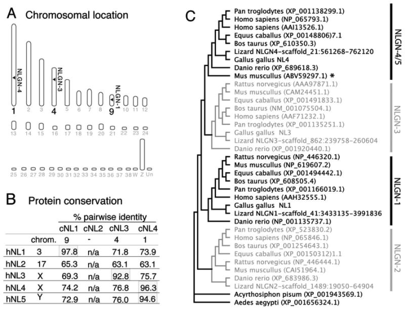
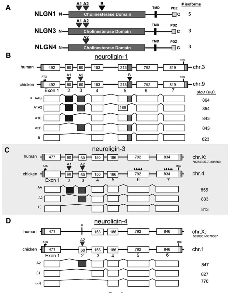
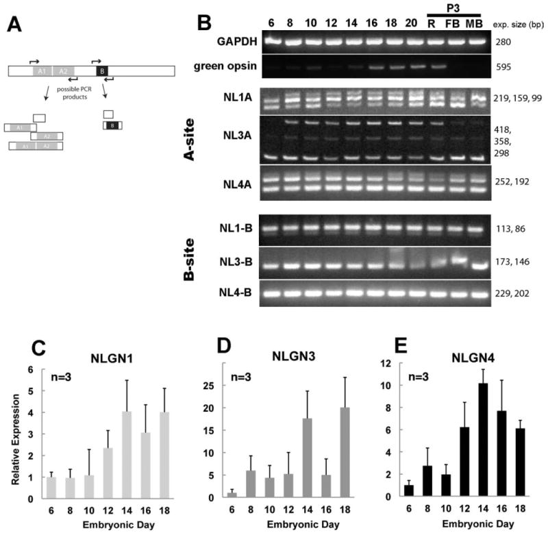
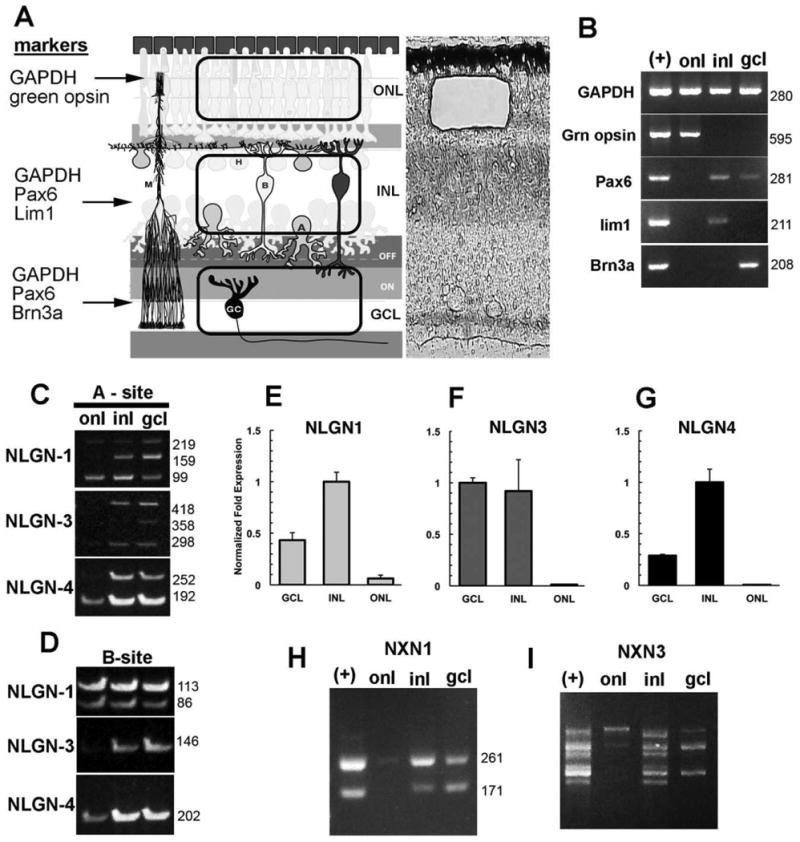
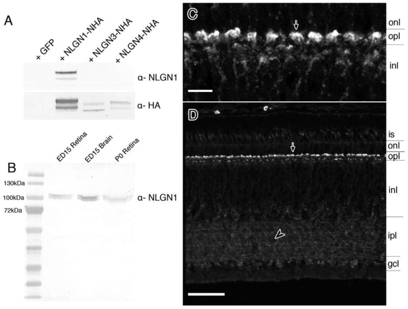
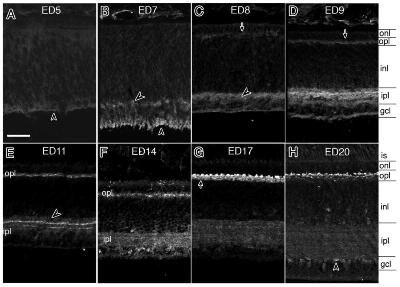
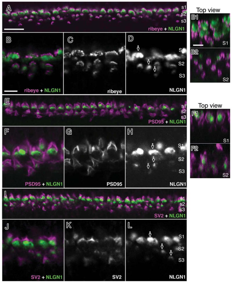
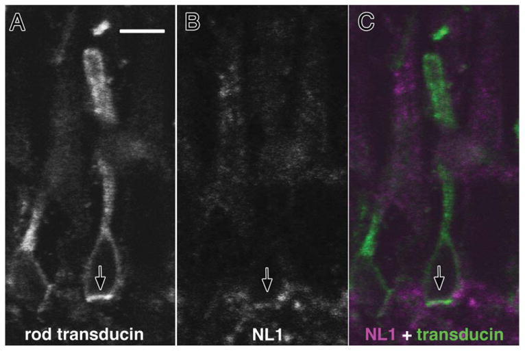
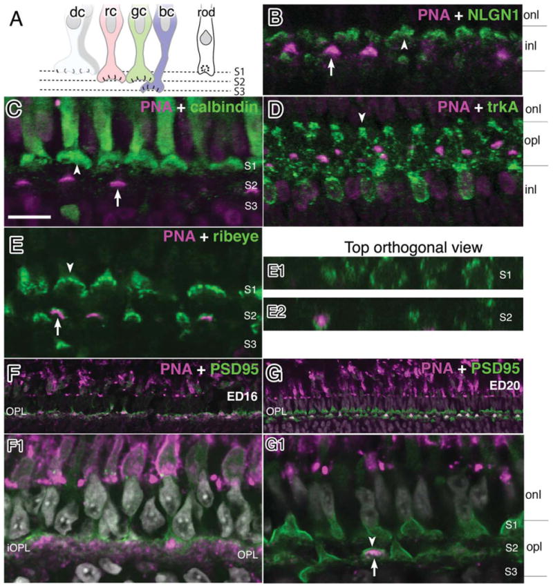
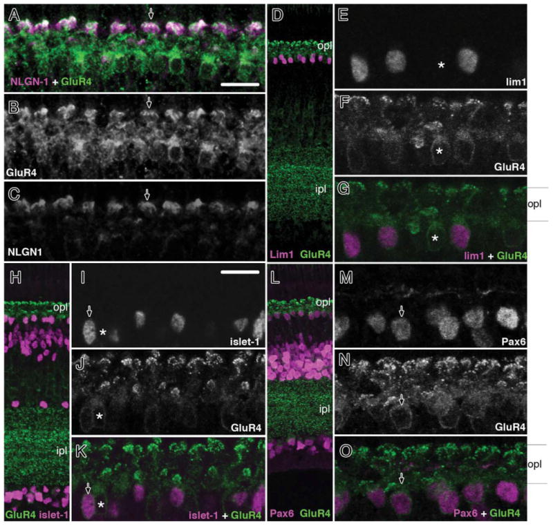
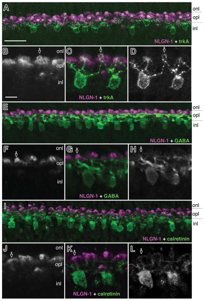
Although synaptogenesis within the retina is obviously essential for vision, mechanisms responsible for the initiation and maintenance of retinal synapses are poorly understood. In addition to its scientific interest, understanding retinal synapse formation is becoming clinically relevant with ongoing efforts to develop transplantation-based approaches for the treatment of retinal degenerative disease. To extend our understanding, we have focused on the chick model system and have studied the neuroligin family of neuronal adhesion factors that has been shown to participate in synapse assembly in the brain. We identified chicken orthologs of neuroligins 1, -3, and -4, but could find no evidence of neuroligin 2. We investigated temporal and spatial patterns of mRNA and protein expression during development using standard polymerase chain reaction (RT-PCR), quantitative PCR (QPCR), laser-capture microdissection (LCM), and confocal microscopy. At the mRNA level, neuroligins were detected at the earliest period tested, embryonic day (ED)5, which precedes the period of inner retina synaptogenesis. Significant alternative splicing was observed through development. While neuroligin gene products were generally detected in the inner retina, low levels of neuroligin 1 mRNA were also detected in the photoreceptor layer. Neuroligin 3 and -4 transcripts, on the other hand, were only detected in the inner retina. At retinal synapses neuroligin 1 protein was detected in the inner plexiform layer, but its highest levels were detected in the outer plexiform layer on the tips of horizontal cell dendrites. This work lays the groundwork for future studies on the functional roles of the neuroligins within the retina.


Figures

Figure 1
Figure 1
Identification and phylogenetic analysis of neuroligin family members. A: There are 38 autosomes and two sex chromosomes (W,Z) in the chicken. The identity of the chromosomes carrying possible NLGN homologs were identified with the Ensembl gene browser and are indicated by horizontal arrowheads. B: Percent identity between the coding regions of human and chicken NLGN genes calculated using a ClustalW alignment shows the degree of homology between the chicken and human orthologs. C: A rooted tree layout cladogram displaying the evolutionary relationships between vertebrate and non-vertebrate animals includes Anolis carolinensis (Lizard), Acyrthosiphon pisum (pea aphid), Aedes aegypti (yellow fever mosquito), Bos taurus (bovine), Danio rerio (zebrafish), Equus caballus (horse), Gallus gallus (chicken), Homo sapiens (human), Mus musculus (mouse), Pan troglodytes (chimpanzee), and Rattus norvegicus (rat). Accession numbers used for alignments and tree building are listed within the dendrogram.
Figure 2
Figure 2
Genomic structure of alternatively spliced isoforms identified in the chick nervous system. A: Cartoon representation of NLGN1, -3, and -4 determined experimentally by sequencing of chicken transcripts from either retina and/or brain. Peptide sequences were aligned to the chicken genome using BLAT. The intron–exon structure of the coding regions for NLGN1 (B), NLGN3 (C), and NLGN4 (D) depict the two major splice sites indicated by inverted red and blue triangles above the A and B splice sites, respectively. The length in basepairs of each exon is indicated within each box and this diagram is not drawn to scale. The amino acid length of each isoform is indicated on the right. The chromosomal positions were included for human NLGN3 and -4 to distinguish between the two genes that are both located on the X-chromosome. The accession numbers used for constructing the human NLGN3 and NLGN4 exon maps were AAF71232.1 and NP_065793.1, respectively. The human NLGN3 did not contain A1 and A2 sites so they were added prior to doing a BLAT alignment. A similar approach was taken for NLGN4. The asterisk above human NLGN4 indicates that NLGN4 is represented in the human genome. The jagged peaks represented by “∧∧∧∧∧∧∧” above coding exons 5 and 6 for chicken NLGN3 signify probable gaps in the current draft of the chicken genome.
Figure 3
Figure 3
Alternatively spliced neuroligin family members expressed during retinal development and relative abundance of gene expression. A: PCR primers flanking each of the conserved splice sites (sites A and B) for NLGN were used to detect NLGN isoform variation and give rise to several combinations of transcript variants. B: PCR amplification of cDNAs from a developmental time course of retinal tissues was separated by gel electrophoresis. C–E: Quantitative real-time PCR shows the relative abundance of NLGN1 (C), NLGN3 (D), and NLGN4 (E) transcripts in relation to the GAPDH housekeeping control gene. Error bars represent standard deviation (n = 3).
Figure 4
Figure 4
Laminar position of alternatively spliced neuroligin and neurexin transcripts in the chick retina. A: The retina is compartmentalized into outer and inner nuclear layers and the ganglion cell layer, each separated by the synaptic plexiform layers. A representative laser capture microdissection cut of the outer nuclear layer is indicated here. B: LCM samples were validated for purity and lack of contamination of RNA from other retinal layers by PCR using primers specific for photoreceptors (green opsin), inner nuclear layer horizontal cells (Lim1), inner retina (Pax6), or the ganglion cells (Brn3). Detection of NLGN1, -3, or -4 alternatively spliced variants in isolated nuclear layers is revealed by analysis with primers flanking splice sites -A (C) or -B (D). E–G: Real-time PCR was used to show relative abundance of NLGN1, -3 and -4 transcripts in relation to the housekeeping gene GAPDH. Different sized alternative spliced NRXN1 (H) and NRXN3 (I) transcripts reveal differences in splicing.
Figure 5
Figure 5
Detection of NLGN1 protein in the chick retina and antibody specificity. A: Western blots with HA-tagged recombinant chicken NLGN1, -3, or -4 proteins were probed with NLGN1 antibodies to demonstrate a lack of cross-reactivity to other NLGN family members. Samples run in parallel were probed with anti-rat HA antibodies as a loading control. B: Native protein from ED15 and P0 chick retinas and ED15 chick forebrain were separated by SDS-PAGE gel electrophoresis and probed with antibodies against NLGN1. C,D: Tissue sections from ED20 retinas were probed with NLGN1 antibodies and detected by fluorescence immunohistochemistry. Panel C is a magnified image of the outer plexiform layer with NLGN1 positive dendrites projecting from the outer inner nuclear layer into the outer plexiform layer (arrow). is, photoreceptor inner segment; onl, outer nuclear layer; opl, outer plexiform; inl, inner nuclear layer; ipl, inner plexiform layer; gcl, ganglion cell layer. Scale bars = 5 μm in C; 25 μm in D.
Figure 6
Figure 6
Developmental patterns of NLGN1 expression during development. A–H: A developmental series of NLGN1 immunoreactivity in the synaptic plexiform layers of the chick retina was carried out from between embryonic day 5 (ED5) and ED20. Arrows indicate preferential detection in the OPL while arrowheads are used to indicate inner plexiform layer features. is, inner segments; onl, outer nuclear layer; opl, outer plexiform layer; sublamina of the outer plexiform layer are indicated as sublayers 1–3 (sublayer1 is located proximal to the onl, sublayer 3 is positioned distal and sublayer 2 is located in between); see text for details. Scale bar = 20 μm.
Figure 7
Figure 7
NLGN1 expression adjacent to photoreceptor terminals in the mature chick retina. Double-labeled immunohistochemistry for NLGN1 and ribeye (A–D), PSD95 (E–H) or SV2 (I–L) is shown in a 21-day-old (ED21) chick retina at the level of the outer plexiform layer. Accumulation of NLGN1 and presynaptic proteins in three distinct strata (S1–3) showed separate labeling patterns between NLGN1 immunoreactive bodies and the boundaries of photoreceptor terminals. Single arrows indicate the ribbed appearance of synaptic ribbon-like structures (D), the peripheral staining of PSD95 (E), or the SV2 positive vesicle pool (I) of double cone terminals. Panels to the right (B1,B2,F1,F2) are top-down orthogonal views showing size differences between synaptic terminals in S1 and S2. Scale bars = 15 μm in A; 5 μm in B,B1.
Figure 8
Figure 8
Neuroligin expression in rod photoreceptors. Double labeling with antibodies against NLGN1 and rod transducin were carried on cryosections from 21-day-old chick retinas. Thin optical sections showing (A) transducin-positive rod photoreceptors were superimposed with (B) NLGN1-positive signals to reveal (C) positive signals at the base of rod photoreceptor terminals. Arrows indicate identical regions of the tissue section across different fluorescent filter sets. Scale bar = 10 μm.
Figure 9
Figure 9
Neuroligin expression in specific sublayers of the OPL. Double labeling immunohistochemistry was performed with antibodies against NLGN1 and markers known to localize to different OPL strata in 21-day-old (ED21) chick retinas. A: A cartoon diagram of photoreceptor projections illustrates the projections of different photoreceptor types that project to different OPL strata. B: AlexaFluor conjugated PNA labeling (magenta) in the medial OPL is indicated with an inverted arrow while NLGN1 signal is in multiple strata (green). C: Calbindin (+) double cone terminals are located in the outer aspect of the OPL (arrowhead) while PNA is located in the medial aspect of the OPL (arrow). D: TrkA positive dendrites extending to the outer OPL terminate near the base of double cones (arrowhead). E: Ribeye signals (arrow) are stratified in all three sublamina with PNA reactivity (arrowhead) restricted to the medial position. Panels to the right (E1,E2) are top-down orthogonal views showing size differences between synaptic terminals in S1 and S2, respectively, and show PNA-negative terminals in S1 and positive PNA terminals in S2 in a number of terminals. A single row of synaptic terminals (arrowhead) is present at ED16 (F,F1). At ED21, three sublaminae (arrowheads) are present (G,G1). dc, double cones; rc, red cones; gc, green cones; bc, blue or violet cones. Scale bar = 10 μm.
Figure 10
Figure 10
NLGN1 and GluR4 expression in dendrites of retinal horizontal cell subpopulations. Double labeling of embryonic day 21 chick retinas (ED21) with NLGN and GluR4 (A–C) or GluR4 and lim1 (D–G), islet1 (H–K), and Pax6 (L–O) showed strong NLGN labeling in different retinal horizontal cell subgroups. Arrowhead (A–C) indicates dendritic tips of double-labeled NLGN1 and GluR4-positive cells. Arrows (G,K,O) indicate GluR4/lim1, GluR4/islet1, and GluR4/Pax6-positive horizontal cells. A single asterisk indicates a horizontal cell that is positive for GluR4 but negative for the above transcription factors. onl, outer nuclear layer; opl, outer plexiform; inl, inner nuclear layer; ipl, inner plexiform layer; gcl, ganglion cell layer. Scale bars = 10 μm.
Figure 11
Figure 11
Neuroligin 1 expression in horizontal cell subsets. Double labeling of embryonic day 21 chick retinas (ED21) with NLGN1 and trkA (A–D), GABA (E–H) or calretinin (I–L) showed colocalization with markers for different retinal horizontal cell subgroups. Arrows indicate NLGN1-positive dendritic tips of horizontal cells that are also positive for trkA(C), GABA(G), and calretinin(K). onl, outer nuclear layer; opl, outer plexiform; inl, inner nuclear layer; ipl, inner plexiform layer; gcl, ganglion cell layer. Scale bars = 15 μm in A; 5 μm in B.

Similar articles

Cited by

References

    1. Aartsen WM, Arsanto JP, Chauvin JP, Vos RM, Versteeg I, Cardozo BN, Bivic AL, Wijnholds J. PSD95beta regulates plasma membrane Ca2+ pump localization at the photoreceptor synapse. Mol Cell Neurosci. 2009;41:156–165. - PubMed
    1. Adler R. Curing blindness with stem cells: hope, reality, and challenges. Adv Exp Med Biol. 2008;613:3–20. - PubMed
    1. Adler R, Lindsey JD, Elsner CL. Expression of cone-like properties by chick embryo neural retina cells in glial-free monolayer cultures. J Cell Biol. 1984;99:1173–1178. - PMC - PubMed
    1. Ashery-Padan R, Marquardt T, Zhou X, Gruss P. Pax6 activity in the lens primordium is required for lens formation and for correct placement of a single retina in the eye. Genes Dev. 2000;14:2701–2711. - PMC - PubMed
    1. Béïque J, Lin D, Kang M, Aizawa H, Takamiya K, Huganir R. Synapse-specific regulation of AMPA receptor function by PSD-95. Proc Natl Acad Sci U S A. 2006;103:19535–19540. - PMC - PubMed

Publication types

MeSH terms