. 2011 Jan;32(1):78-90.
doi: 10.1002/humu.21387.

Dissecting loss of heterozygosity (LOH) in neurofibromatosis type 1-associated neurofibromas: Importance of copy neutral LOH

Affiliations
Free PMC article

Dissecting loss of heterozygosity (LOH) in neurofibromatosis type 1-associated neurofibromas: Importance of copy neutral LOH

Carles Garcia-Linares et al. Hum Mutat. 2011 Jan.
Free PMC article

Abstract

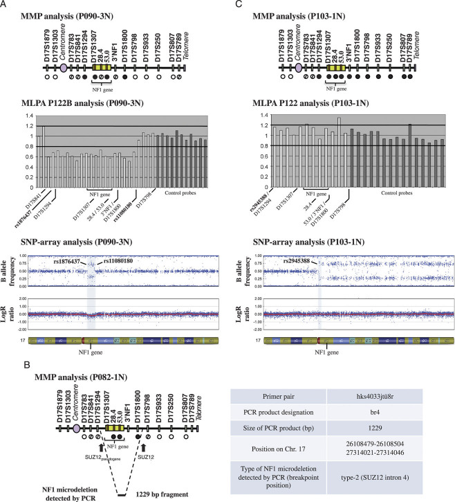
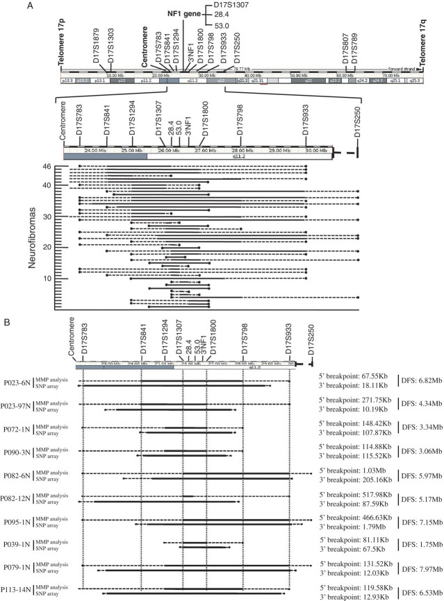
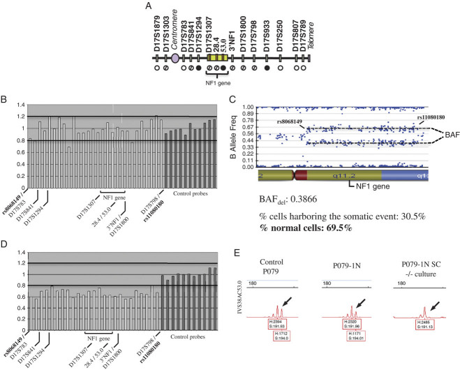
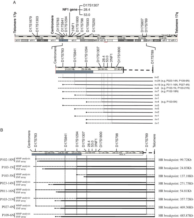
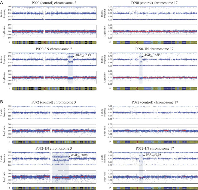
Dermal neurofibromas (dNFs) are benign tumors of the peripheral nervous system typically associated with Neurofibromatosis type 1 (NF1) patients. Genes controlling the integrity of the DNA are likely to influence the number of neurofibromas developed because dNFs are caused by somatic mutational inactivation of the NF1 gene, frequently evidenced by loss of heterozygosity (LOH). We performed a comprehensive analysis of the prevalence and mechanisms of LOH in dNFs. Our study included 518 dNFs from 113 patients. LOH was detected in 25% of the dNFs (N = 129). The most frequent mechanism causing LOH was mitotic recombination, which was observed in 62% of LOH-tumors (N = 80), and which does not reduce the number of NF1 gene copies. All events were generated by a single crossover located between the centromere and the NF1 gene, resulting in isodisomy of 17q. LOH due to the loss of the NF1 gene accounted for a 38% of dNFs with LOH (N = 49), with deletions ranging in size from ∼80 kb to ∼8 Mb within 17q. In one tumor we identified the first example of a neurofibroma-associated second-hit type-2 NF1 deletion. Analysis of the prevalence of mechanisms causing LOH in dNFs in individual patients (possibly under genetic control) will elucidate whether there exist interindividual variation.


Figures

Figure 1
Figure 1
Mechanisms leading to LOH in neurofibromas. A: Mechanisms generating deletions. In all cases Microsatellite Multiplex PCR (MMP) analysis evidenced LOH in the NF1 gene (NG_009018.1) and normally also in 5′ and 3′ regions adjacent to it. MLPA detected only one copy of the NF1 gene. SNP-array analysis detected LOH (four-band pattern in the B allele frequency plot) and one copy of the NF1 gene (LogR ratio <0). B: Nonallelic homologous recombination causing deletion. MMP detected LOH involving the NF1 gene and adjacent regions, apparently not going further 5′ or 3′ of the NF1-REPs. A table summarizing the PCR conditions used for detecting the deletion breakpoint is depicted. Breakpoint localized in SUZ12 intron 4. C: Homologous recombination. MMP evidenced LOH of almost all 17q chromosome. MLPA detected two copies of the NF1 gene and the entire region analyzed. SNP-array detected LOH (four-band pattern in the B allele frequency plot) from the centromere to the end of the chromosome (17q telomere) and the presence of two copies (LogR ratio = 0) of the entire chromosome 17q. MMP (∘ = no LOH; dashed circles = noninformative, • = LOH). MLPA (Values between 0.8 and 1.2 = two copies. Values <0.8 = one copy). SNP-array: B allele frequency plot (0.5 = heterozygote; 0 or 1 = homozygote, other intermediate values = allelic imbalance); LogR ratio (0 = two copies; values <0 = one copy). [Color figures can be viewed in the online issue, which is available at wileyonlinelibrary.com.]
Figure 2
Figure 2
Deletion breakpoint analysis. A: Breakpoint analysis by Multiplex Microsatellite PCR (MMP) and by MLPA analysis. B: Deletion breakpoint mapping refinement by SNP-array. Solid black bar: deletion mapped by either MMP (A, B), by MLPA (A), or SNP-array (B); dashed line: uncertain region by either MMP (A, B) or SNP-array (B). Black circle: no deletion by either MMP (A, B), by MLPA (A) or SNP-array (B). DFS, Deletion fragment size. [Color figures can be viewed in the online issue, which is available at wileyonlinelibrary.com.]
Figure 3
Figure 3
The presence of normal cells in neurofibromas affects the NF1-copy number detection by MLPA. A: MMP analysis of tumor P079-1N (see legend of Fig. 1 for nomenclature); B: MLPA of P079-1N detected two copies of the NF1 gene; C: SNP-array analysis evidenced a high proportion (∼70%) of normal cells within tumor P079-1N; D: MLPA of P079-1N NF1(−/−) Schwann cell culture detected one copy of the NF1 gene. E: Purity of the culture was evidenced by MMP analysis, comparing control, tumor, and SC culture. A total loss of one allele was detected in the SC culture. MLPA (values between 0.8 and 1.2 = two copies. Values <0.8 = one copy). [Color figures can be viewed in the online issue, which is available at wileyonlinelibrary.com.]
Figure 4
Figure 4
Mitotic recombination crossover mapping. A: Crossover analysis by Multiplex Microsatellite PCR (MMP). B: Mapping refinement by SNP-array compared to MMP. Solid black bar: uniparental isodisomy (B); dashed line: uncertain region; black circle: no uniparental isodisomy. HR, Homologous recombination. [Color figures can be viewed in the online issue, which is available at wileyonlinelibrary.com.]
Figure 5
Figure 5
SNP-array analysis of samples P090-3N and P072-1N compared to their respective controls. A: Detection of deletions in 2q and the NF1 region in tumor P090-3N; B: Detection of deletions in 3q and the NF1 region in tumor P072-1N. Deletions were evidenced by a four-band pattern in the B allele frequency plot and a LogR ratio<0.

Similar articles

Cited by

References

    1. Ars E, Serra E, Garcia J, Kruyer H, Gaona A, Lazaro C, Estivill X. Mutations affecting mRNA splicing are the most common molecular defects in patients with neurofibromatosis type 1. Hum Mol Genet. 2000;9:237–247. - PubMed
    1. Assie G, LaFramboise T, Platzer P, Bertherat J, Stratakis CA, Eng C. SNP arrays in heterogeneous tissue: highly accurate collection of both germline and somatic genetic information from unpaired single tumor samples. Am J Hum Genet. 2008;82:903–915. - PMC - PubMed
    1. Bishop AJ, Schiestl RH. Role of homologous recombination in carcinogenesis. Exp Mol Pathol. 2003;74:94–105. - PubMed
    1. Carey JC, Laub JM, Hall BD. Penetrance and variability in neurofibromatosis: a genetic study of 60 families. Birth Defects Orig Artic Ser. 1979;15:271–281. - PubMed
    1. Cavenee WK, Dryja TP, Phillips RA, Benedict WF, Godbout R, Gallie BL, Murphree AL, Strong LC, White RL. Expression of recessive alleles by chromosomal mechanisms in retinoblastoma. Nature. 1983;305:779–784. - PubMed

Publication types