. 2010 Oct 29:8:134.
doi: 10.1186/1741-7007-8-134.

The nucleotide addition cycle of RNA polymerase is controlled by two molecular hinges in the Bridge Helix domain

Affiliations

The nucleotide addition cycle of RNA polymerase is controlled by two molecular hinges in the Bridge Helix domain

Robert O J Weinzierl. BMC Biol. .

Abstract

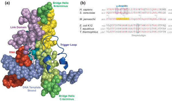
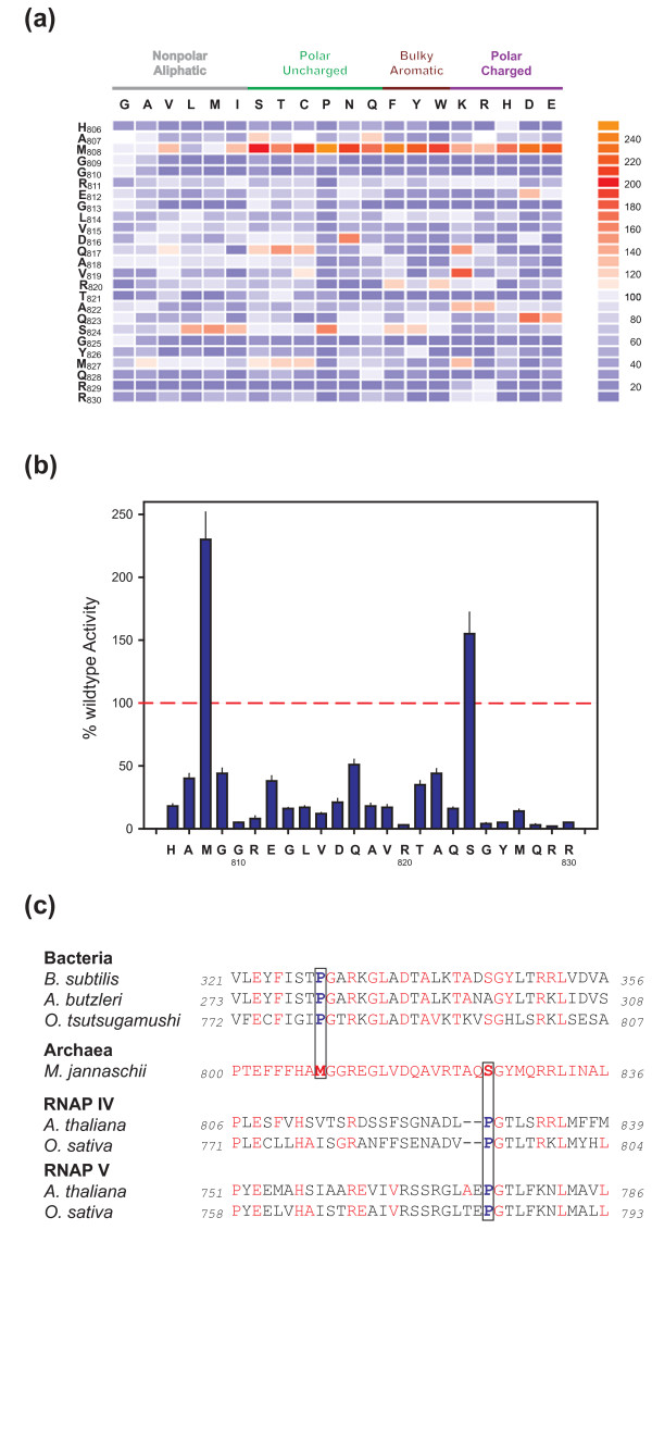
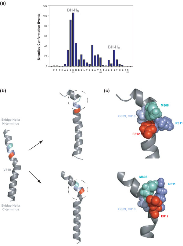
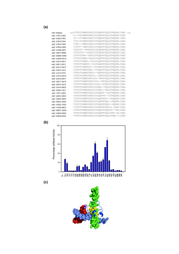
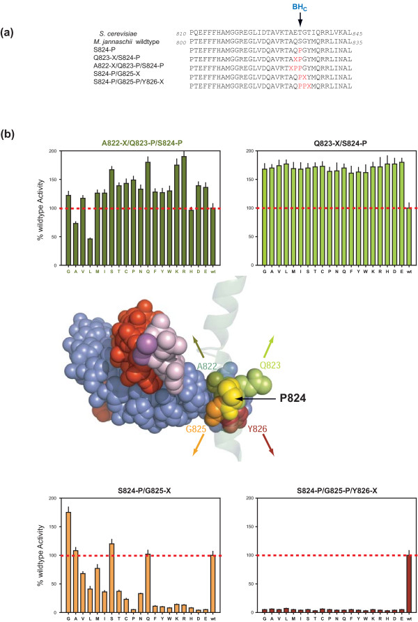
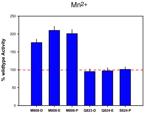
Background: Cellular RNA polymerases (RNAPs) are complex molecular machines that combine catalysis with concerted conformational changes in the active center. Previous work showed that kinking of a hinge region near the C-terminus of the Bridge Helix (BH-H(C)) plays a critical role in controlling the catalytic rate.

Results: Here, new evidence for the existence of an additional hinge region in the amino-terminal portion of the Bridge Helix domain (BH-H(N)) is presented. The nanomechanical properties of BH-H(N) emerge as a direct consequence of the highly conserved primary amino acid sequence. Mutations that are predicted to influence its flexibility cause corresponding changes in the rate of the nucleotide addition cycle (NAC). BH-H(N) displays functional properties that are distinct from BH-H(C), suggesting that conformational changes in the Bridge Helix control the NAC via two independent mechanisms.

Conclusions: The properties of two distinct molecular hinges in the Bridge Helix of RNAP determine the functional contribution of this domain to key stages of the NAC by coordinating conformational changes in surrounding domains.


Figures

Figure 1
Figure 1
Evolutionary conservation and arrangement of domains in the RNAP active site. (A) Most structures are shown in space-filling mode to emphasize spatial connections. The Bridge Helix is shown in green, with the regions subjected to high-throughput mutagenesis in this study highlighted in yellow. The template DNA is pale blue, the RNA is red, the NTP in the insertion site is shown as a pink stick model and catalytic metal ions as magenta spheres. Three additional domains that surround the Bridge Helix N-terminus, β-D II domain (turquoise) and Link (light purple) and the Trigger Loop (dark blue cartoon) are shown. (PDB #2E2H). (B) Alignment of Bridge Helix sequences from bacteria (Escherichia coli K12 (Genbank BAE77332); Thermus aquaticus (Genbank RPOC_THEAQ); Thermus thermophilus (Genbank RPOC_THET8) and eukaryotes (Saccharomyces cerevisiae (Genbank CAA26904) and Homo sapiens (Genbank EAW90183)) against the archaeon M. jannaschii (Genbank A64430). Residues identical to the archaeal sequence are shown in red. The region mutagenized is highlighted with yellow background. The residues contacted by α-amanitin (eukaryotic RNAPII) and streptolydigin (bacterial RNAPs) are boxed. The numbers flanking the sequences represent the location of the sequences within the open reading frame of the complete subunit.
Figure 2
Figure 2
High-throughput mutagenesis of the Bridge Helix. (A) The specific activities in recruitment-independent transcription assays of systematic substitutions of archaeal Bridge Helix residues H806 to R830 are shown as a heat map relative to the activity of the wildtype enzyme (see adjacent scale for comparison). Substitutions in M808 cause an exceptional increase in catalytic activity with chemically diverse side chains. Previously published data (L814 to R830; [20]) are included to provide context. All assays were performed in at least quadruplicate with standard deviations within 12% of the average value. (B) Plot of proline substitutions across the Bridge Helix. The wildtype activity level (100%) is marked with a dashed red line. The substituted residues in the M. jannaschii Bridge Helix are shown along the horizontal axis. Most proline substitutions cause a severe reduction in the specific activity, except at positions M808 and S824, where proline substitutions cause superactivity. All assays were performed in at least quadruplicate, with error bars showing standard deviation from the average value. (C) Naturally occurring proline-substitutions (highlighted in boxes). The Bridge Helix sequences of three bacterial species, Orientia tsutsugamushi (Genbank YP_001248195 (Boryong)/YP_001938485 (Ikeda)), isolates of Arcobacter butzleri (Genbank AAZ80810) and Bacillus subtilis (Genbank BAA10999), as well as representative examples of plant RNAP IV and V Bridge Helix sequences from Arabidopsis thaliana (Genbank AAY89363 and NP_181532, respectively) and Oryza sativa (Genbank EEE70198 and EEE56320, respectively) are aligned against the M. jannaschii sequence (Genbank A64430). The bacterial sequences each contain a single proline residue corresponding to mjA' M808, whereas proline substitutions in RNAP IV and V align with mjA' S824. Residues identical to the archaeal sequence are shown in red.
Figure 3
Figure 3
Structural basis of BH-HN kinking. (A) Overview of local unfolding events incurred by a M. jannaschii Bridge Helix model during 27 independent 200 picosecond MD simulations. The number of 5 picosecond windows during which a specific part of the Bridge Helix adopts a 'coil' conformation are plotted against the Bridge Helix sequence shown on the horizontal axis. A major area of α-helical instability, BH-HN (centered on mjA'G810), is evident from this semi-quantitative analysis. Additional unstable regions include BH-HC (centered on mjA' G825) and a 'labile region' (spanning mjA'Q817 to R820; see Discussion for more details). (B) Examples of kinked BH-HN conformations arising from MD simulations. The Bridge Helix on the left represents the starting conformation as modelled on the yeast RNAPII structure PDB #2E2H. The minor bulge near the center of the Bridge Helix corresponds to the V819 position. During unrestrained simulation, different types of kinked BH-HN structures (circled) involving mjA' G809/G810 (pale blue) and M808 (pale green), R811 (blue) and E812 (red) side-chain interaction emerge stochastically. (C) Structural details of BH-HN kinking models. The relevant residues are shown in space-filling mode to illustrate spatial relationships. Two glycine residues, mjA' G809 and G810 (pale blue) form a highly flexible hinge that allows M808 (pale green) to interact extensively via van der Waals interactions with the side-chains of R811 (blue) and E812 (red). In some cases these interactions create a stretched 310 helix immediately C-terminal to R811 and E812.
Figure 4
Figure 4
Creation of locally twisted Bridge Helix structures. (A) Sequences of the deletion constructs. A two-amino acid deletion window is moved systematically in a single residue step through the entire Bridge Helix primary sequence. The deletions remove two adjacent residues, but, more importantly, join the sequences bordering the mutation with 180° twist because of the removal of half an α-helical turn. (B) Activity of Bridge Helix mutants containing two-amino acid deletions shown relative to wildtype activity (100%). The amino acid pairs deleted from the primary sequence are shown vertically along the horizontal axis. Two distinct peaks of relative insensitivity to the deletions centered on mjA' ΔD816/Q817 and mjA' ΔA822/Q823 are discernible. All assays were performed in at least quadruplicate, with error bars showing standard deviation from the average value. (C) Position of the deletion-insensitive region of the Bridge Helix relative to other elements of the catalytic site. The Bridge Helix and other structures are shown using the same color-scheme as used in Figure 1A (yeast RNAPII elongation complex; [PDB #2E2H]). Residues orthologous to residues displaying the highest activity (>50%) levels in the two-amino acid scan (mjA' D816, Q817, T821, A822 and Q823) are highlighted in yellow.
Figure 5
Figure 5
Functional dissection of the BH-HC region. (A) Position of mutagenized residues. The residues that differ from the archaeal wildtype sequence in the various mutagenesis constructs are shown in red. Wildtype sequences of the M. jannaschii and S. cerevisiae Bridge Helix sequences are shown for comparison. The position of BH-HC is marked with an arrow. (B) The center of the panel provides an overview depicting the orthologous residues in space-filling mode within the mutagenized part of the Bridge Helix modelled on the yeast RNAPII structure (PDB #2E2H); yeast residues E823 and T824 were replaced in silico with Q and P, respectively, to reveal the approximate location of these amino acids relative to the DNA-RNA hybrid and catalytic site). Adjacent parts of the Bridge Helix domain are shown as a transparent ribbon. All other colors are coded as in Figure 1A (template DNA is pale blue, the nascent transcript is red, the NTP in the insertion site is pink and catalytic metal ions are shown as magenta spheres). All mutants shown here contain the mjA' S824-P substitution (yellow). The results of promoter-independent activity assays are plotted relative to wildtype activity (indicated with a red dashed line). Two of the bar charts show the effect of introducing systematic substitutions in positions located immediately N- or C-terminal to S824-P (mjA' Q823 [lime green; upper right]; mjA' G825 [orange; lower left]). The other two bar charts show the functional consequences of introducing additional systematic substitutions in positions located immediately N-terminal to the double proline substitution Q823-P/S824-P (mjA' A822 [olive green; upper left]), or immediately C-terminal to the double proline substitution S824-P/G825 (mjA' Y826 [brown; lower left]). The colors of the histogram bars match the colors of the substituted residues in the structural model. All assays were performed in at least quadruplicate, with error bars showing standard deviation from the average value.
Figure 6
Figure 6
Differential effect of Mn2+ on superactivity in the BH-HN and BH-HC Region. The activities of the most superactive substitutions in BH-HN and BH-HC are compared in Mn2+ substituted reactions relative to the wildtype enzyme. The BH-HN substitutions (mjA' M808-D, -E and -P) continue to display a high degree of superactivity, whereas BH-HC substitutions (mjA' Q823-D, -E and S824-P) only transcribe at rates comparable to wildtype RNAP.
Figure 7
Figure 7
Evolutionary positions of bacterial species with proline-containing Bridge Helices. The evolutionary relationship between three bacterial species with proline-containing Bridge Helices is shown on a rooted phylogenetic tree calculated using maximum-likelihood methods from a concatenation of representative protein sequences [63]. The three species occupy widely divergent branches, strongly suggesting that the proline substitutions evolved independently, rather than were derived from a recently shared ancestor.

Similar articles

Cited by

References

    1. Brueckner F, Ortiz J, Cramer P. A movie of the RNA polymerase nucleotide addition cycle. Curr Opin Struct Biol. 2009;19:294–299. doi: 10.1016/j.sbi.2009.04.005. - DOI - PubMed
    1. Erie DA, Kennedy SR. Forks, pincers, and triggers: the tools for nucleotide incorporation and translocation in multi-subunit RNA polymerases. Curr Opin Struct Biol. 2009;19:708–714. doi: 10.1016/j.sbi.2009.10.008. - DOI - PMC - PubMed
    1. Svetlov V, Nudler E. Macromolecular micromovements: how RNA polymerase translocates. Curr Opin Struct Biol. 2009;19:701–707. doi: 10.1016/j.sbi.2009.10.002. - DOI - PMC - PubMed
    1. Kireeva M, Kashlev M, Burton ZF. Translocation by multi-subunit RNA polymerases. Biochim Biophys Acta. 2010;1799:389–401. - PubMed
    1. Zhang G, Campbell EA, Minakhin L, Richter C, Severinov K, Darst SA. A crystal structure of Thermus aquaticus core RNA polymerase at 3.3 Å resolution. Cell. 1999;98:811–824. doi: 10.1016/S0092-8674(00)81515-9. - DOI - PubMed

Publication types

Substances