. 2010 Oct 29:5:44.
doi: 10.1186/1750-1326-5-44.

Systemic treatment with liver X receptor agonists raises apolipoprotein E, cholesterol, and amyloid-β peptides in the cerebral spinal fluid of rats

Affiliations

Systemic treatment with liver X receptor agonists raises apolipoprotein E, cholesterol, and amyloid-β peptides in the cerebral spinal fluid of rats

Sokreine Suon et al. Mol Neurodegener. .

Abstract

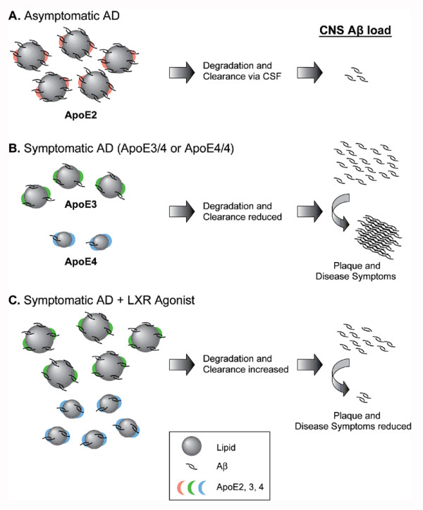
Background: Apolipoprotein E (apoE) is a major cholesterol transport protein found in association with brain amyloid from Alzheimer's disease (AD) patients and the ε4 allele of apoE is a genetic risk factor for AD. Previous studies have shown that apoE forms a stable complex with amyloid β (Aβ) peptides in vitro and that the state of apoE lipidation influences the fate of brain Aβ, i.e., lipid poor apoE promotes Aβ aggregation/deposition while fully lipidated apoE favors Aβ degradation/clearance. In the brain, apoE levels and apoE lipidation are regulated by the liver X receptors (LXRs).

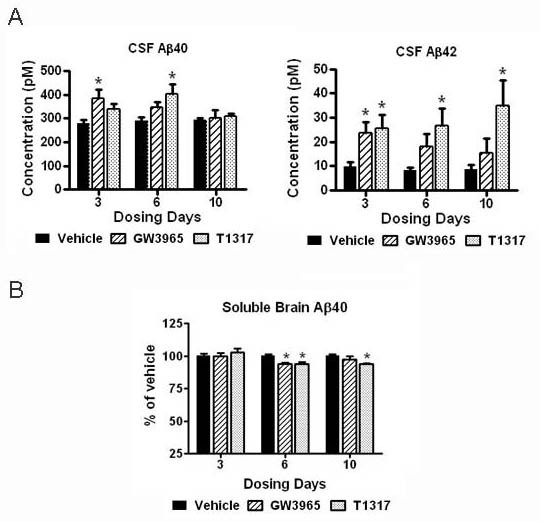
Results: We investigated the hypothesis that increased apoE levels and lipidation induced by LXR agonists facilitates Aβ efflux from the brain to the cerebral spinal fluid (CSF). We also examined if the brain expression of major apoE receptors potentially involved in apoE-mediated Aβ clearance was altered by LXR agonists. ApoE, cholesterol, Aβ40, and Aβ42 levels were all significantly elevated in the CSF of rats after only 3 days of treatment with LXR agonists. A significant reduction in soluble brain Aβ40 levels was also detected after 6 days of LXR agonist treatment.

Conclusions: Our novel findings suggest that central Aβ lowering caused by LXR agonists appears to involve an apoE/cholesterol-mediated transport of Aβ to the CSF and that differences between the apoE isoforms in mediating this clearance pathway may explain why individuals carrying one or two copies of APOE ε4 have increased risk for AD.


Figures

Figure 1
Figure 1
CSF and brain apoE levels were markedly increased with LXR agonist treatment. The levels of apoE in the CSF, soluble brain extracts, and plasma following systemic treatment with LXR agonists T1317 and GW3965 at 30 mpk for 3, 6, and 10 days were evaluated by Western blot analysis as described in Methods. A: Representative blots showing changes in apoE levels in the CSF of rats treated with vehicle or LXR agonists (left panel). Albumin was used as a loading control for all CSF blots. Densitometric analysis of Western blots for CSF apoE (right panel). B: DEA extracted soluble brain homogenates were prepared as described in Methods. Equal amounts of supernatant proteins were loaded per lane and apoJ was used as an additional loading control. Representative blots showing changes in apoE observed in the soluble brain fraction of rats treated with vehicle, T1317 and GW3965 (left panel). Densitometric analysis of Western blots for soluble brain apoE (right panel). C: Representative blots showing changes of apoE levels in the plasma of rats treated with vehicle or LXR agonists (left panel). Densitometric analysis of Western blots for plasma apoE (right panel). * p < 0.05 compared to vehicle treated controls by ANOVA, N = 4 per group.
Figure 2
Figure 2
CSF and soluble brain cholesterol were inversely altered by LXR agonist treatment. The levels of cholesterol in CSF and soluble brain extracts were evaluated using the Amplex Red Cholesterol Kit as described in Methods. A: Cholesterol levels in the CSF of rats treated with LXR agonists in comparison to the vehicle control. B: Cholesterol levels in the DEA-soluble brain fraction of rats treated with LXR agonists in comparison to the vehicle control. * p < 0.05 compared to vehicle treated controls by ANOVA, N = 4 per group. C: CSF samples were separated in native gel as described in Methods and then blotted with an anti-apoE antibody. Representative blot showing apoE immunoreactive bands in the CSF of rats treated with vehicle or T1317 for 3 days. CSF from vehicle-treated rats shows apoE-immunoreactive bands around 240 and 400 kD (←), while CSF from T1317-treated animals had strongest apoE-immunoreactivity in bands detected at approximately 400 and 650 kD (◀).
Figure 3
Figure 3
CSF Aβ levels were increased and soluble brain Aβ levels were decreased by LXR agonists. The levels of Aβ40 and Aβ42 in the CSF and soluble brain extracts were evaluated by ELISA as described in Methods. A: Aβ40 and Aβ42 levels in the CSF of rats treated with LXR agonists in comparison to the vehicle control. B: Aβ40 levels in the DEA-soluble brain fraction of rats treated with LXR agonists in comparison to the vehicle control. * p < 0.05 compared to vehicle treated controls by ANOVA, N = 4 per group.
Figure 4
Figure 4
Brain ABCA1 levels were upregulated, while LDLR and VLDLR were slightly reduced by LXR agonist treatment. The brain levels of the LXR target gene ABCA1 and of the apoE receptors LDLR and VLDLR were evaluated in RIPA extracted brain homogenates by Western blot analysis as described in Methods. Equal amounts of supernatant proteins were loaded per lane and actin was used as an additional loading control. A: Representative blots showing ABCA1 levels in the brain of rats treated with vehicle or LXR agonists (left panel). Densitometric analysis of Western blots for ABCA1 (right panel). B: Representative blots showing LDLR and VLDLR steady-state levels in the brain of rats treated with vehicle, T1317, and GW3965 for 10 days (left panel). Densitometric analysis of Western blots for LDLR and VLDLR (right panel). * p < 0.05 compared to vehicle treated controls by ANOVA, N = 4 per group.
Figure 5
Figure 5
ABCA1 and apoE mRNA expression levels were upregulated in the mouse brain by LXR agonist treatment. ApoE and ABCA1 mRNA as well as protein expression were examined in the mouse brain by Western blot analysis and in situ hybridization following GW3965 treatment (50 mpk for 10 days). A: RIPA and DEA-soluble brain homogenates were prepared as described in Methods. Equal amounts of supernatant proteins were loaded per lane and actin or ApoJ was used as an additional loading control. Densitometric analysis of Western blots for ABCA1 (left panel) and apoE (right panel) levels in the brain of mice treated with vehicle or GW3965. * p < 0.05 compared to vehicle treated controls by ANOVA, N = 9 per group. B: Brain distribution of apoE mRNA by in situ hybridization showing predominant astrocytic expression and increased expression following treatment with GW3965. C: Marked increase of apoE mRNA was observed in the ependymal cells lining the lateral ventricle following LXR agonist treatment. D: In situ hybridization for ABCA1 mRNA also showing high up-regulation in the lateral ventricle by GW3965 (←).
Figure 6
Figure 6
LXR agonist-stimulated cholesterol efflux is apoE- and ABCA1- dependent. The role of ABCA1 in the T1317-induced cholesterol efflux to exogenous apoE was examined in the CCF-STTG1 human astrocytoma cell line. A: HEK 293 cells were transiently transfected with human apoE3 cDNA (E3) or empty vector (V) and concentrated serum-free conditioned media was analysed on Western blotting for apoE content against human recombinant apoE3. B: Equal amounts of conditioned media protein containing approximately 50 μg/ml of HEK-apoE3 were added to CCF-STTG1 astrocytoma cells pre-loaded with cholesterol in serum-free media containing 5 μM T1317 or 0.05% DMSO (vehicle control). 48 h-conditioned media was then analyzed for total cholesterol and protein content while cell monolayers were lysed for Western blotting analysis of ABCA1 protein levels. Treatment with T1317 (5 μM) up-regulated the expression of ABCA1 and increased the release of cholesterol to HEK-apoE3, but not to vector-HEK media. C: Knock-down of ABCA1 with specific siRNA inhibited T1317-induced ABCA1 up-regulation and cholesterol release to HEK-apoE3 media. *p < 0.05 and **p < 0.01 by ANOVA.
Figure 7
Figure 7
Proposed mechanism by which LXR agonists improve apoE-mediated Aβ clearance from the brain. Based on the differential ability of the apoE isoforms to associate with lipids, Aβ, and LDLR, the model predicts that the "protective" apoE2, which has poor binding to LDLR and therefore increased association with lipids and ability to bind Aβ, favors elimination of amyloid to the CSF reducing amyloid deposition within the brain parenchyma (A). In contrast, apoE4, which has decreased association with lipids and reduced Aβ binding capacity when compared to apoE3 and apoE2, can lead to impaired Aβ clearance, increased Aβ oligomerization and deposition into amyloid plaques (B). By raising the levels and the lipidation of apoE and potentially decreasing apoE receptors within the brain, LXR agonists favor the transport of apoE and amyloid to the CSF with subsequent decrease in brain amyloid burden, similar to the "apoE2 effect" (C).

Similar articles

Cited by

References

    1. Selkoe DJ. The molecular pathology of Alzheimer's disease. Neuron. 1991;6:487–498. doi: 10.1016/0896-6273(91)90052-2. - DOI - PubMed
    1. Hardy J, Selkoe DJ. The amyloid hypothesis of Alzheimer's disease: progress and problems on the road to therapeutics. Science. 2002;297:353–356. doi: 10.1126/science.1072994. - DOI - PubMed
    1. Strittmatter WJ, Saunders AM, Schmechel D, Pericak-Vance M, Enghild J, Salvesen GS, Roses AD. Apolipoprotein E: high-avidity binding to beta-amyloid and increased frequency of type 4 allele in late-onset familial Alzheimer disease. Proc Natl Acad Sci USA. 1993;90:1977–1981. doi: 10.1073/pnas.90.5.1977. - DOI - PMC - PubMed
    1. Mahley RW. Apolipoprotein E: cholesterol transport protein with expanding role in cell biology. Science. 1988;240:622–630. doi: 10.1126/science.3283935. - DOI - PubMed
    1. Walker LC, Pahnke J, Madauss M, Vogelgesang S, Pahnke A, Herbst EW, Stausske D, Walther R, Kessler C, Warzok RW. Apolipoprotein E4 promotes the early deposition of Abeta42 and then Abeta40 in the elderly. Acta Neuropathol. 2000;100:36–42. doi: 10.1007/s004010051190. - DOI - PubMed

LinkOut - more resources