. 2010 Oct 29:8:25.
doi: 10.1186/1477-3155-8-25.

Manufacture of IRDye800CW-coupled Fe3O4 nanoparticles and their applications in cell labeling and in vivo imaging

Affiliations

Manufacture of IRDye800CW-coupled Fe3O4 nanoparticles and their applications in cell labeling and in vivo imaging

Yong Hou et al. J Nanobiotechnology. .

Abstract

Background: In recent years, near-infrared fluorescence (NIRF)-labeled iron nanoparticles have been synthesized and applied in a number of applications, including the labeling of human cells for monitoring the engraftment process, imaging tumors, sensoring the in vivo molecular environment surrounding nanoparticles and tracing their in vivo biodistribution. These studies demonstrate that NIRF-labeled iron nanoparticles provide an efficient probe for cell labeling. Furthermore, the in vivo imaging studies show excellent performance of the NIR fluorophores. However, there is a limited selection of NIRF-labeled iron nanoparticles with an optimal wavelength for imaging around 800 nm, where tissue autofluorescence is minimal. Therefore, it is necessary to develop additional alternative NIRF-labeled iron nanoparticles for application in this area.

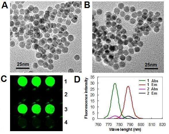
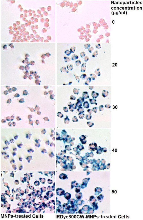
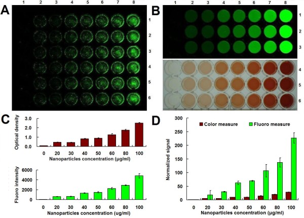
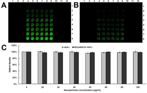
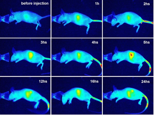
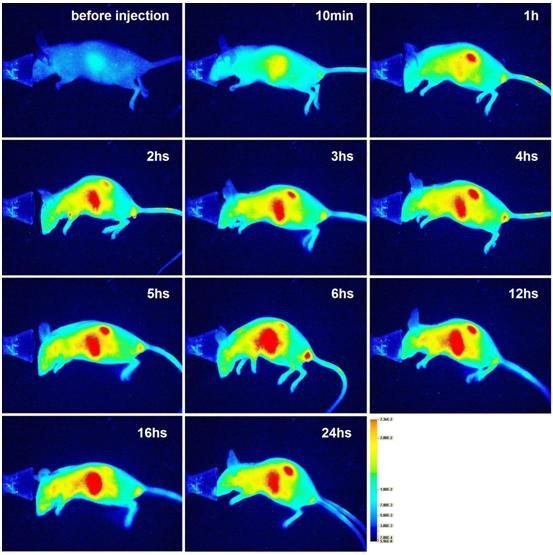
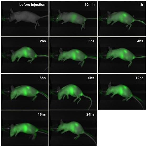
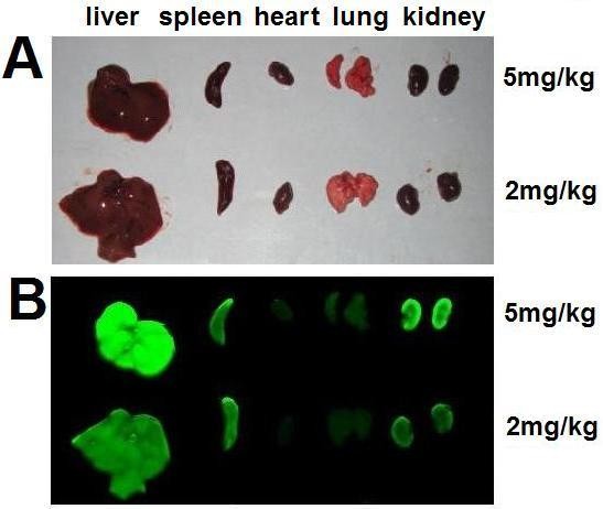
Results: This study manufactured 12-nm DMSA-coated Fe3O4 nanoparticles labeled with a near-infrared fluorophore, IRDye800CW (excitation/emission, 774/789 nm), to investigate their applicability in cell labeling and in vivo imaging. The mouse macrophage RAW264.7 was labeled with IRDye800CW-labeled Fe3O4 nanoparticles at concentrations of 20, 30, 40, 50, 60, 80 and 100 μg/ml for 24 h. The results revealed that the cells were efficiently labeled by the nanoparticles, without any significant effect on cell viability. The nanoparticles were injected into the mouse via the tail vein, at dosages of 2 or 5 mg/kg body weight, and the mouse was discontinuously imaged for 24 h. The results demonstrated that the nanoparticles gradually accumulated in liver and kidney regions following injection, reaching maximum concentrations at 6 h post-injection, following which they were gradually removed from these regions. After tracing the nanoparticles throughout the body it was revealed that they mainly distributed in three organs, the liver, spleen and kidney. Real-time live-body imaging effectively reported the dynamic process of the biodistribution and clearance of the nanoparticles in vivo.

Conclusion: IRDye800CW-labeled Fe3O4 nanoparticles provide an effective probe for cell-labeling and in vivo imaging.


Figures

Figure 1
Figure 1
Characterization of IRDy800CW-MNPs. (A) TEM image of MNPs. (B) TEM image of IRDy800CW-MNPs. (C) NIRF signal of nanoparticles. (D) Fluorescent spectrum of the nanoparticles. 1: IRDy800CW-MNPs; 2: MNPs. 3-4: The resuspended precipitate and supernatant of the IRDy800CW-MNPs solution after heat treatment and centrifugation. Abs: absorbance. Em: emission.
Figure 2
Figure 2
Prussian blue staining of cells. The agglomerates of Fe3O4 nanoparticles are stained in blue.
Figure 3
Figure 3
Measurement of the relative iron-loading of cells. (A) NIRF signal of cells labeled with IRDy800CW-MNPs at doses of 0, 20, 30, 40, 50, 60, 80 and 100 μg/ml (Column 1-8). Each dose contained 6 repeats (Row 1-6). Cells were washed with PBS before imaging. (B) Measurement of the relative iron-loading of cells (A) with colorimetric and NIRF approaches. Row 1-3: NIRF signals; Row 4-6: Colorimetric signals. (C) The intensity of colorimetric and NIRF signals (B). (D) The normalized intensity of colorimetric and NIRF signals (C). The signal of the nanoparticle-labeled cells was normalized to that of the negative control cell. The error bars represent mean and standard deviations of experiments performed in triplicate.
Figure 4
Figure 4
Measurement of cell viability. (A) NIRF signals of cells treated with MNPs (Column 1-3 and 10-12) and IRDy800CW-MNPs (Column 4-9) at doses of 0, 20, 30, 40, 50, 60, 80 and 100 μg/ml (From row 1-8) for 24 h. (B) NIRF signals of cells (A) after washing three times with PBS. (C) Quantitative measurement of cell viability by MTT assay. The error bars represent mean and standard deviations of experiments performed with 6 repeats.
Figure 5
Figure 5
NIRF imaging of a mouse administered IRDye800CW-MNPs at a dose of 2 mg/kg body weight. The images are displayed in pseudo-color mode.
Figure 6
Figure 6
NIRF imaging of a mouse administered the IRDy800CW-MNPs at a dose of 5 mg/kg body weight. The images are displayed in pseudo-color mode.
Figure 7
Figure 7
NIRF imaging of a mouse administered the IRDye800CW-MNPs at a dose of 5 mg/kg body weight. The images are displayed in an overlay mode of light channel image and NIRF channel image.
Figure 8
Figure 8
NIRF imaging of organs of a IRDy800CW-MNPs-injected mouse. (A) Light images of heart, lung, liver, spleen and kidney of the mouse administered the IRDye800CW-MNPs. (B) NIRF images of the same organs.

Similar articles

Cited by

References

    1. Lewin M, Carlesso N, Tung CH, Tang XW, Cory D, Scadden DT, Weissleder R. Tat peptide-derivatized magnetic nanoparticles allow in vivo tracking and recovery of progenitor cells. Nat Biotechnol. 2000;18:410–414. doi: 10.1038/74464. - DOI - PubMed
    1. Nam JM, Thaxton CS, Mirkin CA. Nanoparticle-based bio-bar codes for the ultrasensitive detection of proteins. Science. 2003;301:1884–1886. doi: 10.1126/science.1088755. - DOI - PubMed
    1. Weissleder R, Moore A, Mahmood U, Bhorade R, Benveniste H, Chiocca EA, Basilion JP. In vivo magnetic resonance imaging of transgene expression. Nat Med. 2000;6:351–355. doi: 10.1038/73219. - DOI - PubMed
    1. Tiefenauer L. In: Nanotechnology in Biology and Medicine. Tuan Vo-Dinh, editor. Boca Raton: CRC Press; 2007. Magnetic Nanoparticles as Contrast Agents for Medical Diagnosis. 29/10.
    1. Ito A, Shinkai M, Honda H, Kobayashi T. Heat-inducible TNF-alpha gene therapy combined with hyperthermia using magnetic nanoparticles as a novel tumor-targeted therapy. Cancer Gene Ther. 2001;8:649–654. doi: 10.1038/sj.cgt.7700357. - DOI - PubMed