. 2010 Nov 12;87(5):593-603.
doi: 10.1016/j.ajhg.2010.10.001. Epub 2010 Oct 28.

Prodynorphin mutations cause the neurodegenerative disorder spinocerebellar ataxia type 23

Affiliations

Prodynorphin mutations cause the neurodegenerative disorder spinocerebellar ataxia type 23

Georgy Bakalkin et al. Am J Hum Genet. .

Erratum in

  • Am J Hum Genet. 2010 Nov 12;87(5):736

Abstract

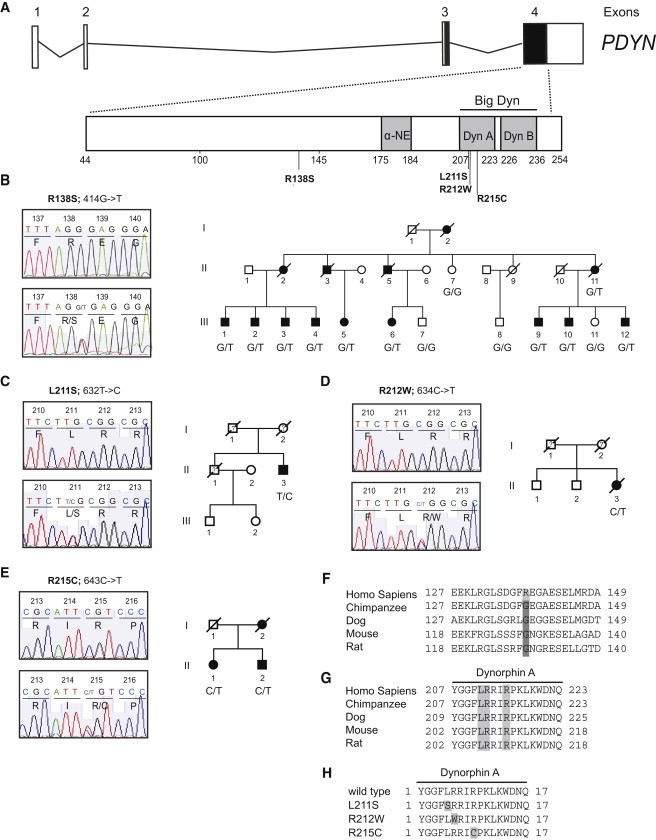
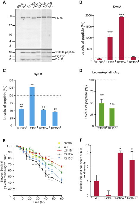
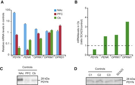
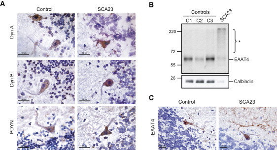
Spinocerebellar ataxias (SCAs) are dominantly inherited neurodegenerative disorders characterized by progressive cerebellar ataxia and dysarthria. We have identified missense mutations in prodynorphin (PDYN) that cause SCA23 in four Dutch families displaying progressive gait and limb ataxia. PDYN is the precursor protein for the opioid neuropeptides, α-neoendorphin, and dynorphins A and B (Dyn A and B). Dynorphins regulate pain processing and modulate the rewarding effects of addictive substances. Three mutations were located in Dyn A, a peptide with both opioid activities and nonopioid neurodegenerative actions. Two of these mutations resulted in excessive generation of Dyn A in a cellular model system. In addition, two of the mutant Dyn A peptides induced toxicity above that of wild-type Dyn A in cultured striatal neurons. The fourth mutation was located in the nonopioid PDYN domain and was associated with altered expression of components of the opioid and glutamate system, as evident from analysis of SCA23 autopsy tissue. Thus, alterations in Dyn A activities and/or impairment of secretory pathways by mutant PDYN may lead to glutamate neurotoxicity, which underlies Purkinje cell degeneration and ataxia. PDYN mutations are identified in a small subset of ataxia families, indicating that SCA23 is an infrequent SCA type (∼0.5%) in the Netherlands and suggesting further genetic SCA heterogeneity.


Figures

Figure 1
Figure 1
PDYN Mutations Causing SCA23 (A) PDYN exons 3 and 4 encode PDYN, which gives rise to the opioid peptides α-neoendorphin (α-NE), dynorphin A (Dyn A), dynorphin B (Dyn B), and big dynorphin (Big Dyn), which encompasses Dyn A and Dyn B. DNA sequence analysis identified four missense mutations (c.414G>T, c.632T>C, c.634C>T, and c.643C>T) in SCA23 subjects. (B) Sequence electropherogram and pedigree of original Dutch SCA23 family. The c.414G>T; p.R138S mutation was identified in 10 affected individuals, but not in the four unaffected family members. (C–E) Sequence electropherograms and pedigrees of the patients with Dyn A mutations: c.632T>C; p.L211S, c.634C>T; p.R212W, and c.643C>T; p.R215C. At the moment, no DNA material of additional family members was available for mutation screening in the p.L211S and p.R212W families. The p.R215C mutation was identified in two affected siblings. Closed symbols denote affected; open symbols denote unaffected; ? denotes disease status unknown; / denotes deceased. (F and G) Conservation of the mutated amino acids. Arginine 138 is human specific; other species analyzed have glycine 138. Leucine 211, Arginine 212, and Arginine 215 are conserved across species. (H) Localization of mutations in Dyn A.
Figure 2
Figure 2
SCA23 Mutations Affect PDYN Processing and Enhance Dyn A Toxicity (A) Expression and processing of WT and mutant PDYNs in RINm-5F cells. Immunoblotting was performed with anti-PDYN (against C-terminal fragment; top) or anti-Dyn B (bottom) antibodies. Synthetic big dynorphin (a 32 amino acid peptide consisting of Dyn A and Dyn B) and Dyn B were loaded on the same gel as peptide markers and produced the 4 and 2 kDa bands identified by anti-Dyn B antibodies (data not shown; however, their positions are shown on the right). The 10, 4, and 2 kDa PDYN processing products were detected in RINm-5F cells transfected with PDYN expression plasmids. (B–D) Peptide levels in cells expressing WT PDYN were taken as 100%. One-way ANOVA followed by Dunnett's multiple comparison test; ∗∗p < 0.01, ∗∗∗p < 0.001; mutant versus wild-type. Data are shown as means ± SEM. (B) RINm-5F cells expressing PDYN p.L211S and p.R212W showed significant elevation in levels of Dyn A compared to cells expressing WT PDYN (ANOVA; p < 0.001). The Dyn A RIA was not hindered by the mutations because they did not affect the binding of the mutant peptides to the Dyn A antibodies. The antibodies were generated against the C-terminal Dyn A fragment.12,19 (C) Dyn B levels showed significant decrease in cells expressing PDYN p.R138S, p.R212W, and p.R215C (ANOVA; p < 0.001). (D) PDYN p.R138S and p.R215C produced lower levels of Leu-enkephalin-Arg compared to WT PDYN (ANOVA; p < 0.005). Leu-enkephalin-Arg peptides derived from PDYN p.L211S and p.R212W were not analyzed by RIA because these two mutations are located within the Leu-enkephalin-Arg antigenic epitope and may hinder the binding of these mutant peptides to anti-Leu-enkephalin-Arg antibodies.12,19 (E and F) Time-lapse imaging of striatial neurons treated with Dyn A peptides (100 nM) revealed marked loss of neuronal viability induced by Dyn A p.R212W and Dyn A p.R215C peptides compared to vehicle-treated control or WT Dyn A (one-way repeated-measures ANOVA; p < 0.005). Data are represented as the means ± SEM. (F) Neuronal death at 60 hr induced by mutant Dyn A peptides. The level of neuronal death as a result of excess WT Dyn A was considered as 1. Bonferroni's multiple correction test, p < 0.05. Data are represented as the means ± SD.
Figure 3
Figure 3
Impact of the PDYN p.R138S Mutation on Expression of the Opioid Genes and PDYN Levels in Human Brain (A) Levels of opioid mRNA in the nucleus accumbens (NAc), prefrontal cortex (PFC), and cerebellum (Cb) were quantified by TaqMan low-density arrays in postmortem samples from three control subjects. PDYN expression was substantially lower in Cb compared to NAc and PFC, the two brain areas with high and moderate gene expression. In the Cb, PENK showed high expression levels compared to the PFC but lower expression levels compared to the NAc. Both OPRK1 and OPRM1 are expressed in Cb, NAc, and PFC at similar levels, whereas OPRD1 expression was not detectable in the Cb. Data bars represent the means ± SEM. (B) Comparison of mRNA levels in the Cb between three control and SCA23 cerebella. The data is shown as the ratio of levels in SCA23 to those in controls. Mean levels in controls were set to 1, as indicated by the dashed line. PENK, OPRK1, and OPRM1 expression was 2- and 3.5-fold increased, whereas PDYN expression was slightly decreased in the SCA23 Cb compared to controls. (C) Analysis of PDYN in the human NAc, PFC, and Cb by immunoblot analysis. Tissue samples were pooled from three control subjects and purified on SEP-PAC reverse-phase columns prior to SDS-PAGE. PDYN levels in cerebellum are intermediate between those in NAc and PFC. (D) No differences were evident between PDYN levels in control (C1-3) and SCA23 cerebella.
Figure 4
Figure 4
Localization of PDYN and Dynorphins, and Analysis of the Purkinje Cell-Specific Excitatory Amino Acid Transporter, in SCA23 Cerebellum (A) Immunohistochemistry of cerebellum from the PDYN p.R138S subject and a control individual without neurological disease. Immunoperoxidase labeling with anti-Dyn A, anti-Dyn B, or anti-PDYN antibodies was visualized by 3-Amino-9-ethylcarbazole as the chromogen and was counterstained with hematoxylin. (B) Immunoblot analysis of excitatory amino acid transporter (EAAT4) and calbindin, both Purkinje cell markers. High molecular weight species were detected in the SCA23 cerebellum (). (C) Immunoperoxidase labeling of EAAT4 in SCA23 and control cerebellum. Scale bars represent 30 μm (A and C).

Similar articles

Cited by

References

    1. Schöls L., Amoiridis G., Büttner T., Przuntek H., Epplen J.T., Riess O. Autosomal dominant cerebellar ataxia: Phenotypic differences in genetically defined subtypes? Ann. Neurol. 1997;42:924–932. - PubMed
    1. Matilla-Dueñas A., Sánchez I., Corral-Juan M., Dávalos A., Alvarez R., Latorre P. Cellular and molecular pathways triggering neurodegeneration in the spinocerebellar ataxias. Cerebellum. 2010;9:148–166. - PubMed
    1. Sato N., Amino T., Kobayashi K., Asakawa S., Ishiguro T., Tsunemi T., Takahashi M., Matsuura T., Flanigan K.M., Iwasaki S. Spinocerebellar ataxia type 31 is associated with “inserted” penta-nucleotide repeats containing (TGGAA)n. Am. J. Hum. Genet. 2009;85:544–557. - PMC - PubMed
    1. Schorge S., van de Leemput J., Singleton A., Houlden H., Hardy J. Human ataxias: A genetic dissection of inositol triphosphate receptor (ITPR1)-dependent signaling. Trends Neurosci. 2010;33:211–219. - PMC - PubMed
    1. Carlson K.M., Andresen J.M., Orr H.T. Emerging pathogenic pathways in the spinocerebellar ataxias. Curr. Opin. Genet. Dev. 2009;19:247–253. - PMC - PubMed

Publication types

LinkOut - more resources