. 2010 Nov 12;40(3):364-76.
doi: 10.1016/j.molcel.2010.10.011. Epub 2010 Oct 28.

Regulation of the histone H4 monomethylase PR-Set7 by CRL4(Cdt2)-mediated PCNA-dependent degradation during DNA damage

Affiliations

Regulation of the histone H4 monomethylase PR-Set7 by CRL4(Cdt2)-mediated PCNA-dependent degradation during DNA damage

Hisanobu Oda et al. Mol Cell. .

Abstract

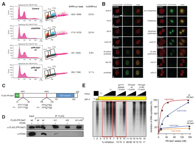
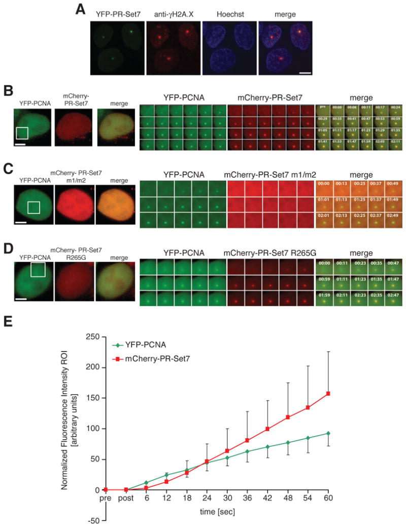
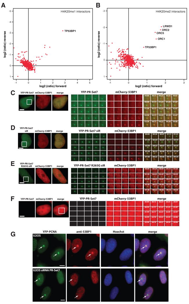
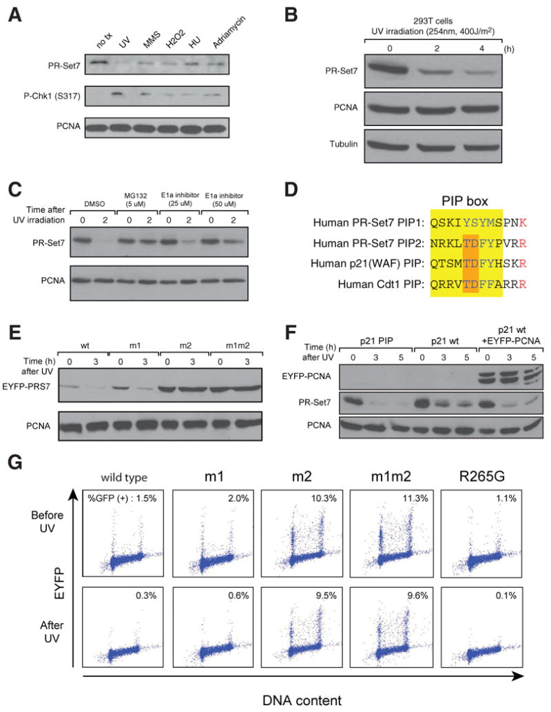
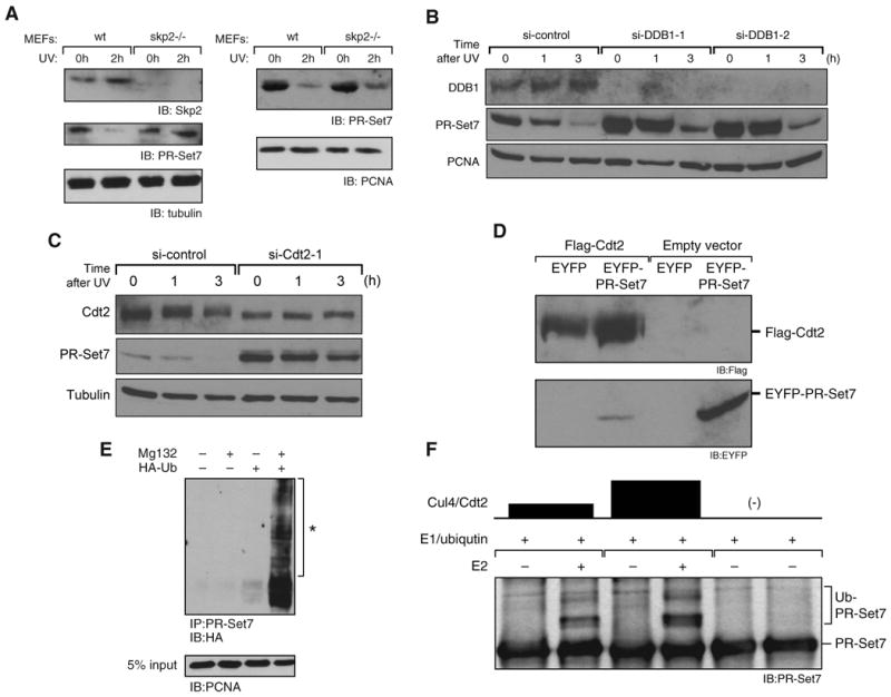
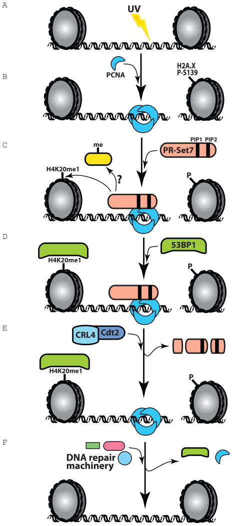
The histone methyltransferase PR-Set7/Set8 is the sole enzyme that catalyzes monomethylation of histone H4 at K20 (H4K20me1). Previous reports document disparate evidence regarding PR-Set7 expression during the cell cycle, the biological relevance of PR-Set7 interaction with PCNA, and its role in the cell. We find that PR-Set7 is indeed undetectable during S phase and instead is detected during late G2, mitosis, and early G1. PR-Set7 is transiently recruited to laser-induced DNA damage sites through its interaction with PCNA, after which 53BP1 is recruited dependent on PR-Set7 catalytic activity. During the DNA damage response, PR-Set7 interaction with PCNA through a specialized "PIP degron" domain targets it for PCNA-coupled CRL4(Cdt2)-dependent proteolysis. PR-Set7 mutant in its "PIP degron" is now detectable during S phase, during which the mutant protein accumulates. Outside the chromatin context, Skp2 promotes PR-Set7 degradation as well. These findings demonstrate a stringent spatiotemporal control of PR-Set7 that is essential for preserving the genomic integrity of mammalian cells.


Figures

Figure 1
Figure 1. PR-Set7 is undetectable in S phase and interacts with PCNA in vitro
(A) FACS analysis of U2OS cells stably expressing EYFP-PR-Set7. The Y-axis shows cell count and fluorescence intensity of EYFP derived from EYFP-PR-Set7. The X-axis represents DNA content. The majority of positive cells were observed at G1 and G2/M, but hardly detectable during S phase. si-RNAs of PR-Set7 reduce the number of cells which have a EYFP signal (bottom two panels). (B) Single cell time-lapse analysis reveals that YFP-PR-Set7 is undetectable during S phase, but detectable from late G2 through mitosis to early G1. S-phase is visualized by mCherry-PCNA labeling of replication foci. YFP-PR-Set7, green; mCherry-PCNA, red; representative data of n=35 cells is shown; scale bar, 5 μm. (C) Schematic of recombinant human PR-Set7 protein used for protein interaction assays. Two PIP domains (PIP1 and PIP2) are depicted with their amino acid sequences shown below; amino acids changed in the PIP mutants m1 and m2 are underlined. N-terminal FLAG-tag and SET domain are shown by rectangles in green and blue, respectively. (D) PR-Set7 interacts with PCNA in a PIP domain dependent manner. Western blot analysis using anti-FLAG immunoprecipitations performed with purified recombinant FLAG-tagged PR-Set7, either wild type or mutant in the PR-Set7 PIP domains, and purified recombinant PCNA. (E) PR-Set7 can inhibit PCNA dependent DNA polymerization in vitro, in a PIP domain dependent manner. PR-Set7, as well as p21, inhibits nucleotide incorporation by DNA polymerase δ in a DNA polymerization assay performed in vitro. The area outlined in red rectangles shows corresponding lanes in which the same incremental amounts of PCNA were used in the assay. On the right: Graph of percentage inhibition of DNA synthesis calculated from the results shown in (E).
Figure 2
Figure 2. PR-Set7 is recruited to DNA damage sites in a PCNA dependent manner
(A) PR-Set7 co-localizes with γ-H2A.X foci at sites of laser-induced DNA damage. YFP-PR-Set7, green; anti-γ-H2A.X immunolabeling, red; Hoechst DNA labeling, blue; representative data of n=21 cells is shown; scale bar, 5 μm. (B) PCNA and PR-Set7 are recruited to DNA damage sites. YFP-PCNA, green; mCherry-PR-Set7, red; time post irradiation in sec; representative data of n=30 cells is shown; scale bar, 5 μm; the enlarged area is indicated by a white square. (C) PIP domains of PR-Set7 are essential for its recruitment to DNA damage sites. YFP-PCNA, green; mCherry-PR-Set7 m1/m2, red; representative data of n=8 cells is shown; scale bar, 5 μm (D) Catalytic activity of PR-Set7 is dispensable for its recruitment to DNA damage sites. YFP-PCNA, green; mCherry-PR-Set7 R265G, red; representative data of n=8 cells is shown; scale bar, 5 μm. (E) Quantification of mCherry-PR-Set7 and YFP-PCNA recruitment to a DNA damage site. Average fluorescence intensities of the ROI over background, arbitrary units; error bars, standard deviation (n=7).
Figure 3
Figure 3. PR-Set7 catalytic activity is required for 53BP1 recruitment to a DNA damage site
(A) 53BP1 binds H4K20me1, as determined by a SILAC-based histone peptide pull-down approach. Proteins are plotted according to their SILAC-ratio in the “forward” (x-axis; unmodified peptide incubated with “light” extract, modified peptide incubated with “heavy” extract) and “reverse” (y-axis; unmodified peptide incubated with “heavy” extract, modified peptide incubated with “light” extract) experiment. Specific interactors should lie close to the diagonal in the upper right quadrant. Background binders cluster together in the bottom left part of the graph, showing a 1:1 ratio in both experiments. (B) 53BP1 binds H4K20me2, as determined by a SILAC-based histone peptide pull-down approach. Plot is as described in (A). (C) PR-Set7 recruitment to a DNA damage site precedes that of 53BP1. YFP-PR-Set7, green; mCherry-53BP1, red; representative data of n=8 cells is shown; scale bar, 5 μm. (D) Cells treated with siRNA against PR-Set7 exhibit similar 53BP1 recruitment to a DNA damage site as in (B), upon transient expression of siRNA resistant WT PR-Set7. YFP-PR-Set7 siR, green; mCherry-53BP1, red; representative data of n=7 cells is shown; scale bar, 5 μm (E) In contrast to (C), siRNA-PR-Set7 treated cells are devoid of detectable 53BP1 recruitment to a DNA damage site upon transient expression of the catalytically inactive PR-Set7 mutant (R265G). YFP-PR-Set7 R265G, green; mCherry-53BP1, red; representative data of n=8 cells is shown; scale bar, 5 μm. (F) PR-Set7 is required for 53BP1 binding to the DNA damage site. Cells treated with PR-Set7 siRNA for 3 days were transfected with mCherry-53BP1. YFP-PR-Set7, green; mCherry-53BP1, red; representative data of n=10 cells is shown; scale bar, 5 μm. (G) Recruitment of endogenous 53BP1 to a DNA damage site requires PR-Set7. U2OS cells expressing YFP-PCNA were mock treated or transfected for 3 days with siRNA against PR-Set7. Cells were fixed 6 min after the laser pulse and subjected to immunofluorescence labeling of endogenous 53BP1. YFP-PCNA, green; anti-53BP1, red; Hoechst DNA labeling, blue; arrows indicate the irradiated site; representative data of n=6 (U2OS) or n=10 (U2OS siRNA PR-Set7) cells is shown; scale bar, 5 μm.
Figure 4
Figure 4. The PIP2 domain is required for PR-Set7 degradation during the cell cycle and after UV irradiation
(A) PR-Set7 protein levels decreased after treating U2OS cells with a panel of DNA damaging agents, as follows: UV irradiation (400 J/m2), MMS (3mM, 1 hr), H2O2 (500uM, 30m), HU (1mM, 16 hr), and Adriamycin (1 μg/ml, 4 hr). Phospho-Chk1-serine 317 served as a marker for DNA damage, and PCNA as a loading control. (B) The protein levels of PR-Set7 decrease after UV irradiation (400 J/m2) in HEK293T cells as analyzed by western blotting. Both PCNA and β-tubulin serve as loading controls. (C) UV-treated HEK293T cells exhibit increased PR-Set7 levels in the presence of proteasome inhibitor or ubiquitin activating enzyme E1 inhibitor. (D) Comparison of PR-Set7 PIP sites with other PIP degron containing proteins. Amino acid residues of the PIP box are highlighted in yellow. The TD residues and a basic amino acid at +4 (red) in the PIP degron are conserved between the PR-Set7 PIP2 and PIP degrons of human p21 and Cdt1. (E) PR-Set7 mutant in its PIP2 domain (m2 or m1/m2) does not exhibit degradation after UV irradiation, relative to PR-Set7 either wild type or mutant in its PIP1 domain (m1). (F) Overexpression of wild type p21, but not its PIP mutant, attenuates PR-Set7 degradation in UV-treated HEK293T cells. PR-Set7 degradation is restored upon co-expressing EYFP-PCNA protein with wild type p21. (G) FACS analysis of cells expressing EYFP-tagged PR-Set7, either wild type or mutant in one (m1 or m2) or both (m1/m2) PIP domains, or catalytically inactive (R265G), as a function of UV-treatment (400 J/m2). Irrespective of UV-treatment, GFP positive cells expressing PR-Set7 mutant in PIP2 are more abundant.
Figure 5
Figure 5. PR-Set7 is degraded by the CRL4Cdt2 ubiquitin ligase
(A) Loss of Skp2 shows partial stabilization of PR-Set7 after UV treatment. PR-Set7 protein levels were analyzed in both Skp2-/- and wild type MEFs after UV treatment. Western blot analysis of the levels of PR-Set7 in whole cell extracts (left side) and in chromatin bound proteins (right side). Tubulin and PCNA serve as loading controls, respectively. (B) siRNA mediated knockdown of DDB1 results in PR-Set7 accumulation irrespective of UV-treatment. Western blot of chromatin fraction derived from non-irradiated or UV-irradiated (400 J/m2) 293T cells after DDB1 knockdown. PCNA served as a loading control. (C) siRNA mediated knockdown of Cdt2 prevents PR-Set7 degradation irrespective of UV treatment. Similar experiment as in (B), using tubulin as a loading control. (D) Transient expression levels of EYFP-PR-Set7 are reduced upon co-transfection with FLAG-Cdt2 in 293T cells. (E) Endogenous PR-Set7 is polyubiquitylated in vivo after treatment with MG132. Western blot analysis using anti-HA antibody of endogenous PR-Set7 immunoprecipitated from 293T cells after transfection with HA-Ubiquitin,. Polyubiquitylated PR-Set7 is indicated with an asterisk. (F) CRL4Cdt2 E3 ligase ubiquitylates PR-Set7. Results from an ubiquitylation assay performed in vitro using affinity purified CRL4Cdt2 and recombinant PR-Set7. Ubiquitin-conjugated PR-Set7 (Ub-PR-Set7).
Figure 6
Figure 6. Model for Regulation of PR-Set7 at DNA Damage sites
(A) DNA damage induces H2A.X phosphorylation, and PCNA gets recruited to the DNA damage site (B) PR-Set7 binds to PCNA through its PIP domains (via interaction through PIP1 and/or PIP2) (C) PR-Set7 methylates H4K20, and possibly other non-histone proteins. (D) 53BP1 binds to monomethylated lysine H4K20 through its double Tudor domains. (E) CRL4Cdt2 ubiquitylates PR-Set7 through the interaction with PIP2, followed by proteasomal degradation. (F) PCNA remains at the DNA damage site and works as a platform to recruit other repair proteins.

Comment in

References

    1. Abbas T, Sivaprasad U, Terai K, Amador V, Pagano M, Dutta A. PCNA-dependent regulation of p21 ubiquitylation and degradation via the CRL4Cdt2 ubiquitin ligase complex. Genes Dev. 2008;22:2496–2506. - PMC - PubMed
    1. Anastassiadis K, Kim J, Daigle N, Sprengel R, Scholer HR, Stewart AF. A predictable ligand regulated expression strategy for stably integrated transgenes in mammalian cells in culture. Gene. 2002;298:159–172. - PubMed
    1. Arias EE, Walter JC. PCNA functions as a molecular platform to trigger Cdt1 destruction and prevent re-replication. Nat Cell Biol. 2006;8:84–90. - PubMed
    1. Botuyan MV, Lee J, Ward IM, Kim JE, Thompson JR, Chen J, Mer G. Structural basis for the methylation state-specific recognition of histone H4-K20 by 53BP1 and Crb2 in DNA repair. Cell. 2006;127:1361–1373. - PMC - PubMed
    1. Dignam JD, Lebovitz RM, Roeder RG. Accurate transcription initiation by RNA polymerase II in a soluble extract from isolated mammalian nuclei. Nucleic Acids Res. 1983;11:1475–1489. - PMC - PubMed

Publication types

MeSH terms