Comment
. 2010 Nov 16;18(5):485-98.
doi: 10.1016/j.ccr.2010.10.002. Epub 2010 Oct 28.

CD4(+) T cells contribute to the remodeling of the microenvironment required for sustained tumor regression upon oncogene inactivation

Affiliations
Comment

CD4(+) T cells contribute to the remodeling of the microenvironment required for sustained tumor regression upon oncogene inactivation

Kavya Rakhra et al. Cancer Cell. .

Erratum in

  • Cancer Cell. 2010 Dec 14;18(6):696

Abstract

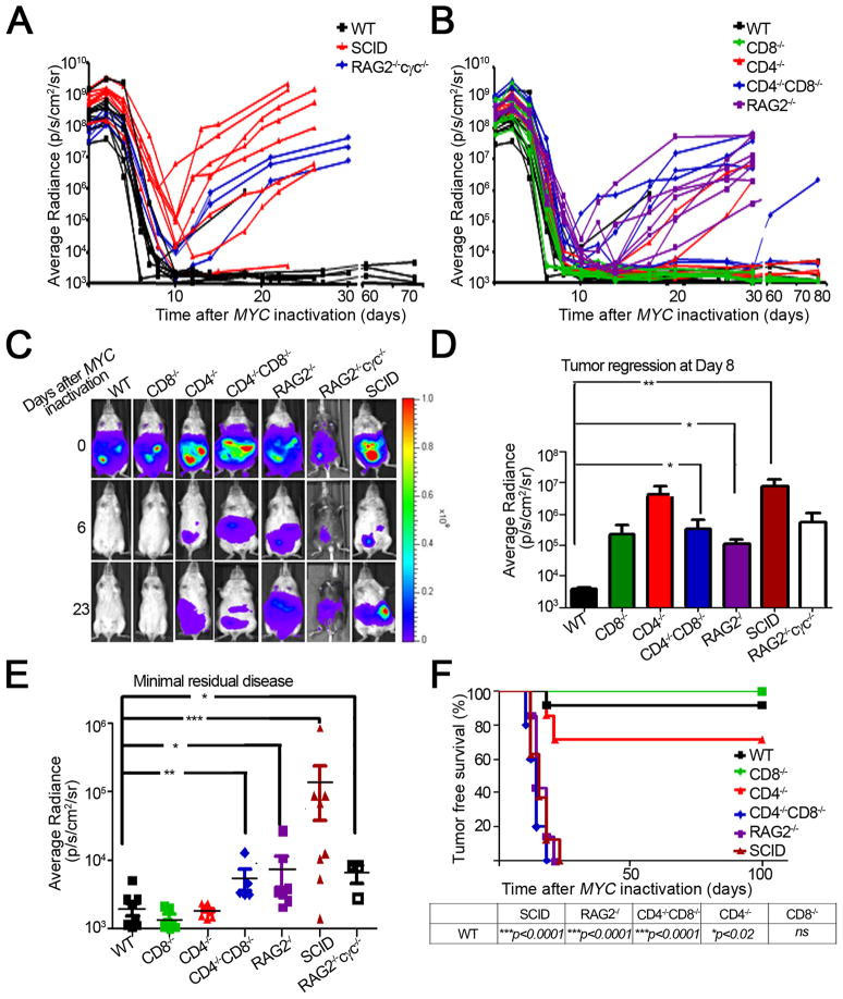
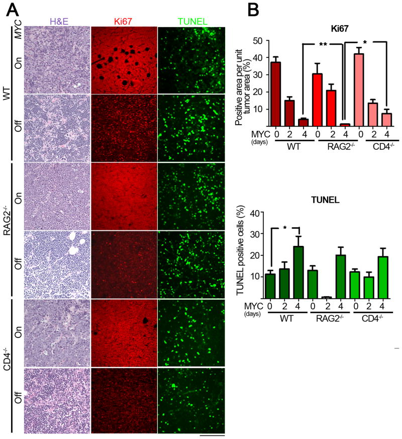
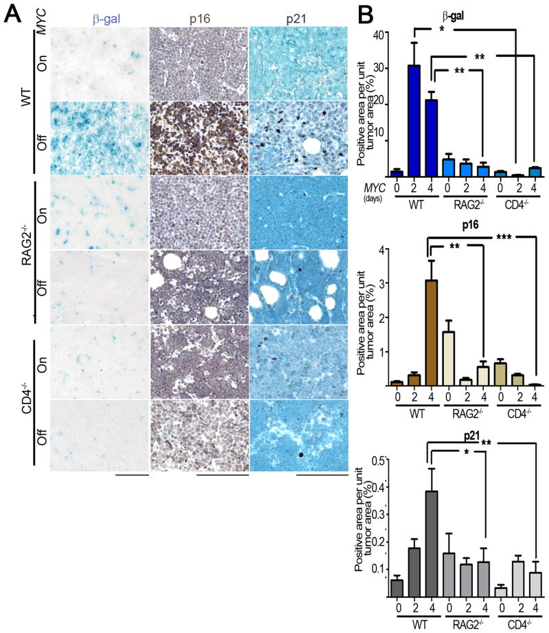
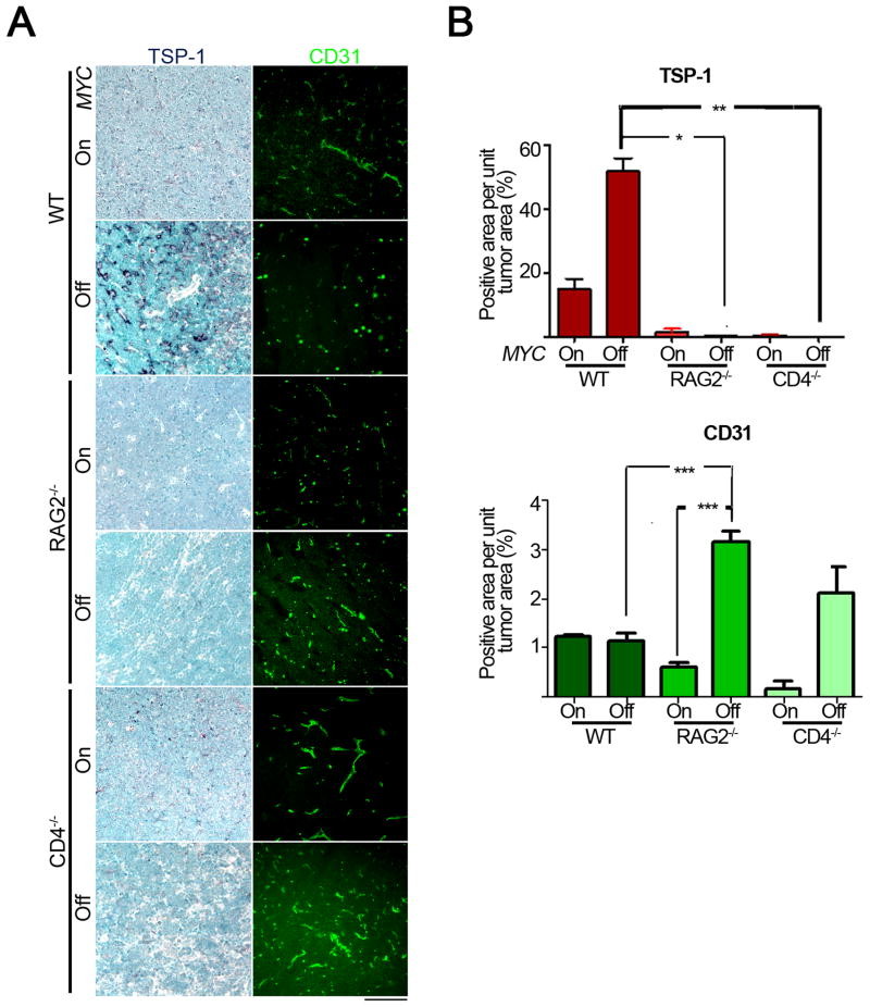
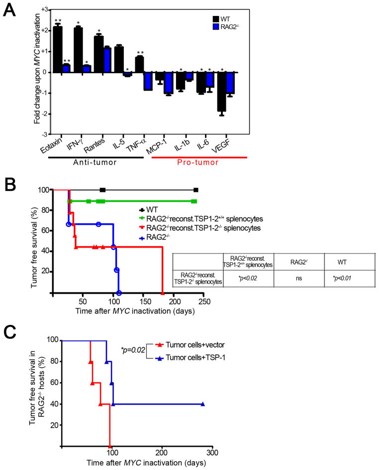
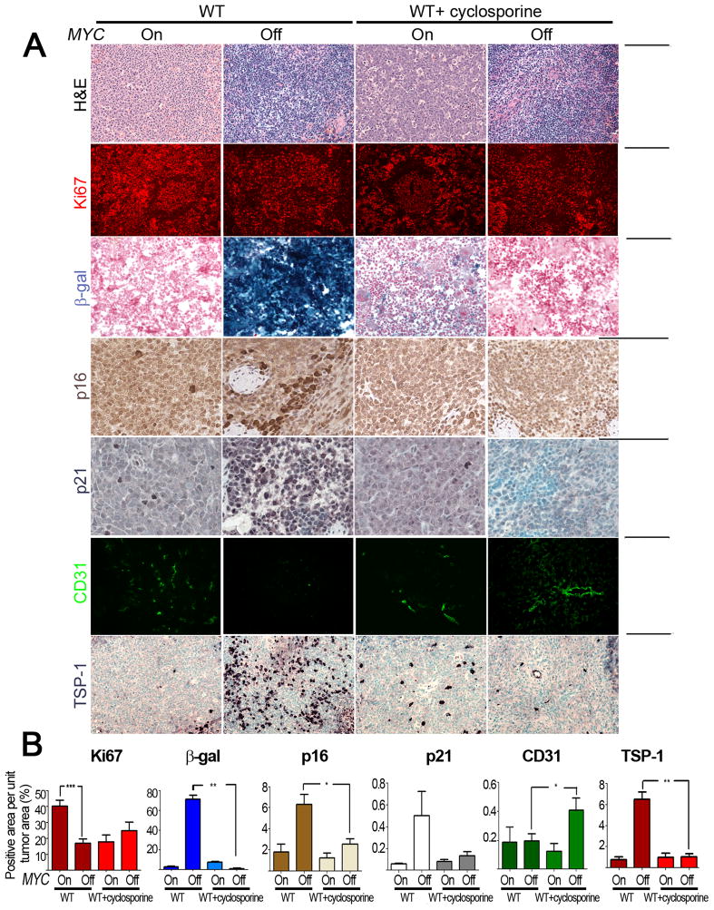
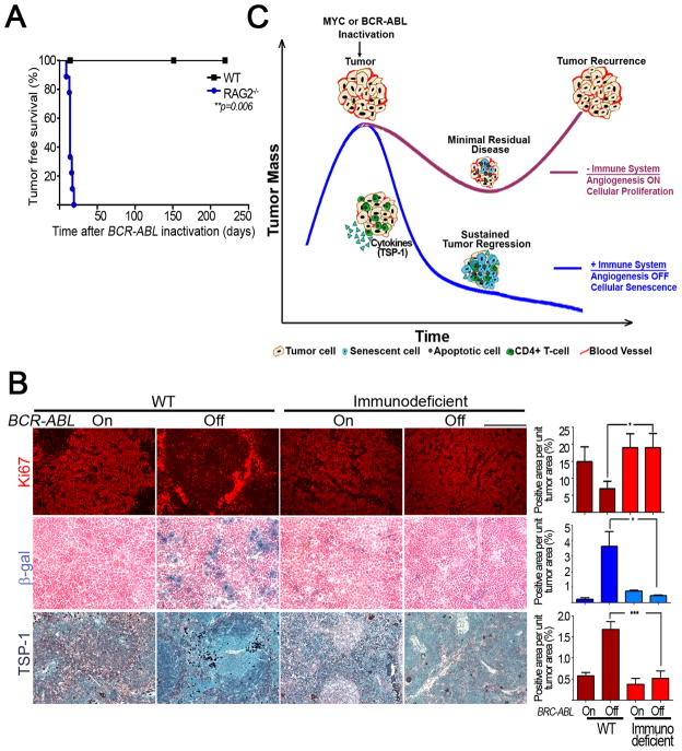
Oncogene addiction is thought to occur cell autonomously. Immune effectors are implicated in the initiation and restraint of tumorigenesis, but their role in oncogene inactivation-mediated tumor regression is unclear. Here, we show that an intact immune system, specifically CD4(+) T cells, is required for the induction of cellular senescence, shutdown of angiogenesis, and chemokine expression resulting in sustained tumor regression upon inactivation of the MYC or BCR-ABL oncogenes in mouse models of T cell acute lymphoblastic lymphoma and pro-B cell leukemia, respectively. Moreover, immune effectors knocked out for thrombospondins failed to induce sustained tumor regression. Hence, CD4(+) T cells are required for the remodeling of the tumor microenvironment through the expression of chemokines, such as thrombospondins, in order to elicit oncogene addiction.


Figures

Figure 1
Figure 1. An intact immune system is required for sustained tumor regression
1(A, B): Graphical representation of tumor regression and relapse kinetics as measured by bioluminescence imaging. Luciferase-labeled tumor cell lines from our conditional mouse T-ALL model were injected s.c. into different cohorts of mice (WT n = 11, CD8−/− n = 7, CD4−/− n = 6, CD4−/−CD8−/− n = 5, SCID n = 8, RAG2−/− n = 7, RAG2−/−cγc−/− n = 3). MYC was inactivated by administering doxycycline (dox) to the mice when tumors reached a comparable bioluminescence signal (108p/s/sr/cm2). 1(C): Bioluminescence images of tumors regressing in the different immunodeficient hosts. Data is representative of 3 experiments. 1(D): Quantitative analysis of tumor regression in the indicated hosts, 8 days post MYC inactivation. 1(E): Quantification of minimum residual disease in the indicated hosts. Data is presented as the minimum bioluminescence signal after MYC inactivation. 1(F): Kaplan Meier curves of tumor-free survival in the various immunodeficient genotypes. A mouse was scored as a relapse when its tumor bioluminescence signal first began to increase after tumor regression. The log-rank test was used to compare survival curves. Data is representative of 3 experiments using 2 cell lines and 1 primary tumor. Statistical significance was analyzed by pooling data from all experiments (WT n = 43, CD8−/− n = 20, CD4−/− n = 24, CD4−/−CD8−/− n = 15, SCID n = 15, RAG2−/− n = 46) (p value evaluated by unpaired Student’s t-test) is shown. * p < 0.01, ** p < 0.001, *** p < 0.0001. Error bars are +/− SEM. See also Figure S1
Figure 2
Figure 2. The immune system does not influence apoptosis or proliferative arrest upon MYC inactivation
2(A): Micrographs of Hematoxylin and Eosin staining (left panel), TUNEL (middle panel) and Ki67 (right panel) immunostaining of tumors derived from untreated (MYC On), two-day and four-day dox treated mice (MYC Off) from WT (top panel) RAG2−/− (middle panel) and CD4−/− (bottom panel) hosts. Scale Bar = 100 μm. 2(B): Quantitative representation of Ki67 (top panel) and TUNEL (bottom panel) immunostaining shown in 2(A) for 0, 2 and 4d after MYC inactivation. Quantification of TUNEL and Ki67 immunostaining is presented as the average percentage of TUNEL-positive cells and area of Ki67-positive regions, respectively, within the tumors. At least five different fields from three different tumors injected with at least two different tumor cell lines for each different condition. Statistical significance (p value evaluated by unpaired Student’s t-test) is shown. * p < 0.01, ** p < 0.001, *** p < 0.0001. Error bars are represented as +/− SEM.
Figure 3
Figure 3. An intact immune system is required for the induction of cellular senescence upon MYC inactivation
3(A): Micrographs of Senescence Associated β-galactosidease (SA β-gal, left panel), p16 (middle panel) and p21 (right panel) immunostaining of tumors derived from untreated (MYC On), two-day and four-day dox treated (MYC Off) mice of the indicated genotypes. Scale Bar = 100 μm. 3(B): Quantification of SA-β-gal (top panel), p16 (middle panel) and p21 (bottom panel) staining shown in 3(A) for 0, 2 and 4 days after MYC inactivation. Quantification is presented as the average percentage of positively stained regions within the tumors. At least five different fields from three different tumors injected with at least two different tumor cell lines were analyzed for each different condition. Statistical significance (p value evaluated by unpaired Student’s t-test) is shown. * p < 0.01, ** p < 0.001, *** p < 0.0001. Error bars are +/− SEM.
Figure 4
Figure 4. An intact immune system is required for the inhibition of angiogenesis upon MYC inactivation
4(A): Micrographs of TSP-1 (left panel) and CD31 (right panel) immunohistochemical and immunofluorescence staining of tumors derived from untreated (MYC On) and 4 day dox treated (MYC Off) mice of the indicated genotypes. Scale Bar = 100μm. 4(B): Quantification of TSP-1 (top panel) and CD31 (bottom panel) staining shown in 4(A). Quantification is presented as the average percentage of positively stained regions within the tumors. At least five different fields from two different tumors were analyzed for each different condition. Statistical significance (p value evaluated by unpaired Student’s t-test) is shown. * p < 0.01, ** p < 0.001, *** p < 0.0001. Error bars are +/− SEM. See also Figure S2
Figure 5
Figure 5. CD4+ T-cells home to the tumor and are sufficient to induce sustained tumor regression upon MYC inactivation
5(A): Bioluminescence signal of luciferase+CD4+ T-cells that home to the tumor microenvironment. RAG2−/− mice were reconstituted with luciferase+ CD4+ T-cells and unlabeled tumor cell lines were injected s.c. 8 days post reconstitution. MYC was inactivated when tumors grew to a size of 1000 mm3. Data is represented as bioluminescence signal (average radiance) plotted against time after MYC inactivation (n=3). 5(B): Tumor regression and relapse kinetics measured by bioluminescence imaging. RAG2−/− mice were reconstituted with CD4+ (RAG2−/−reconst. CD4+Tcells, n=5) or CD8+ (RAG2−/−reconst. CD8+Tcells, n=6) T-cells from WT mice. 8 days after reconstitution, luciferase+ tumor cell lines were injected s.c. MYC was inactivated when tumors in all hosts reached a comparable bioluminescence signal. Data is presented as bioluminescence signal (average radiance) plotted against time after MYC inactivation. WT (n=3) and RAG2−/− (n=3) mice were used as positive and negative controls. 5(C): Quantification of minimum residual disease. Bioluminescence signals of tumors at their maximally regressed state are plotted against genotype. Statistical significance (p value evaluated by unpaired Student’s t-test) is shown. * p < 0.01, ** p < 0.001, *** p < 0.0001. Error bars are +/− SEM. 5(D): Kaplan Meier curves of tumor-free survival in the reconstituted RAG2−/−, RAG2−/− and WT mice. Log-rank test was used compare the survival curves. Data is representative of 3 experiments. Statistics were performed including all data: n = 14, RAG reconstituted with CD8+ T-cells: n = 12. reconst. = reconstituted with. See also Figure S3.
Figure 6
Figure 6. Cytokines produced by the immune system contribute to sustained tumor regression upon MYC inactivation
6(A): Graphical representation of fold change of indicated cytokines upon MYC inactivation in tumors from WT and RAG2−/− hosts. Tumors from WT and RAG2−/− mice were harvested at tumor onset and 4 days after MYC inactivation and run on a luminex platform to check for protein expression of 21 different cytokines. The significant fold changes in the various cytokines upon MYC inactivation were log2 transformed and plotted for various pro- and anti-tumor cytokines. *p < 0.01, ** p < 0.001. * above the bars represents significance in cytokine expression upon MYC inactivation in the indicated host. Error bars are +/− SEM. 6(B): Kaplan-Meier curves of tumor free survival of reconstituted RAG2−/−, RAG2−/− and WT mice. RAG2−/− mice were reconstituted with splenocytes from WT (n=18) or TSP-1,2−/− (n=16) mice i.v. 8 days post reconstitution mice were transplanted with lymphoma cells s.c. MYC was inactivated when tumors were 1000 mm3. WT (n=8) and RAG2−/− (n=11). Log-rank test was used to analyze survival of indicated genotypes. Data is representative 3 experiments. 6(C): Kaplan-Meier curves of tumor free survival of RAG2−/− mice injected with TSP-1 transfected tumor cell lines (n = 5) or vector transfected control tumor cell lines (n = 5). A p53−/− conditional MYC lymphoma cell line was used. See also Figure S4
Figure 7
Figure 7. Cyclosporine A treatment inhibits induction of senescence and inhibition of angiogenesis in tumors from primary MYC induced T-ALL
7(A): Micrographs of Hematoxylin and Eosin, Ki67, SA-β-gal, p16, p21, CD31 and TSP-1 immunostaining (ordered from top to bottom) of tumors derived from untreated and cyclosporine A treated primary tumor bearing mice (MYC On and 4 day dox treated MYC Off). Scale Bar = 100μm. 7(B): Quantification of immunostaining shown in 7(A) above. Ordered from left to right, the graphs represent quantification of Ki67, SA-β-gal, p16, p21, CD31 and TSP-1 expression. Quantification is the average percentage of positively stained regions within the tumors. At least five different fields from two different tumors were analyzed. Statistical significance (p value evaluated by unpaired Student’s t-test) is shown. * p < 0.01, ** p < 0.001, *** p < 0.0001. Error bars are +/− SEM. See also Figure S5.
Figure 8
Figure 8. An intact immune system is required for sustained regression of tumors in a conditional mouse model of BCR-ABL-induced B-ALL
8(A): Kaplan-Meier curves of tumor free survival of RAG2−/− (n=9) and WT (n=4) mice transplanted with unlabelled leukemia cells i.p. When mice were moribund with tumor, BCR-ABL was inactivated, and mice were scored for relapse. 8(B): Micrographs and quantification of Ki67, SA-β-gal and TSP-1 immunostaining (ordered from top to bottom) of tumors derived from untreated (BCR-ABL On) and doxycycline treated (BCR-ABL Off) wildtype and immunodeficient tumor bearing mice. Scale Bar = 100μm. Quantification is average percentage of positively stained regions. At least five different fields from two different tumors were analyzed for each different condition. Statistical significance (p value evaluated by unpaired Student’s t-test) is shown. * p < 0.01, ** p < 0.001, *** p < 0.0001. 8(C): Model for role of immune system in eliciting oncogene addiction. See also Figure S6.

Comment in

Comment on

Similar articles

Cited by

References

    1. Acosta JC, O’Loghlen A, Banito A, Guijarro MV, Augert A, Raguz S, Fumagalli M, Da Costa M, Brown C, Popov N, et al. Chemokine signaling via the CXCR2 receptor reinforces senescence. Cell. 2008;133:1006–1018. - PubMed
    1. Aguirre-Ghiso JA. Models, mechanisms and clinical evidence for cancer dormancy. Nature reviews. 2007;7:834–846. - PMC - PubMed
    1. Allavena P, Sica A, Solinas G, Porta C, Mantovani A. The inflammatory micro-environment in tumor progression: the role of tumor-associated macrophages. Crit Rev Oncol Hematol. 2008;66:1–9. - PubMed
    1. Andreu P, Johansson M, Affara NI, Pucci F, Tan T, Junankar S, Korets L, Lam J, Tawfik D, DeNardo DG, et al. FcRgamma activation regulates inflammation-associated squamous carcinogenesis. Cancer cell. 2010;17:121–134. - PMC - PubMed
    1. Beatty G, Paterson Y. IFN-gamma-dependent inhibition of tumor angiogenesis by tumor-infiltrating CD4+ T cells requires tumor responsiveness to IFN-gamma. J Immunol. 2001;166:2276–2282. - PubMed

Publication types

MeSH terms