Review
. 2010 Dec 10;9(12):1299-306.
doi: 10.1016/j.dnarep.2010.10.001. Epub 2010 Oct 28.

Mre11-Rad50-Nbs1 conformations and the control of sensing, signaling, and effector responses at DNA double-strand breaks

Affiliations
Review

Mre11-Rad50-Nbs1 conformations and the control of sensing, signaling, and effector responses at DNA double-strand breaks

Gareth J Williams et al. DNA Repair (Amst). .

Abstract

Repair and integrity of DNA ends at breaks, replication forks and telomeres are essential for life; yet, paradoxically, these responses are, in many cases, controlled by a single protein complex, Mre11-Rad50-Nbs1 (MRN). The MRN complex consists of dimers of each subunit and this heterohexamer controls key sensing, signaling, regulation, and effector responses to DNA double-strand breaks including ATM activation, homologous recombinational repair, microhomology-mediated end joining and, in some organisms, non-homologous end joining. We propose that this is possible because each MRN subunit can exist in three or more distinct states; thus, the trimer of MRN dimers can exist in a stunning 6(3) or 216 states, a number that can be expanded further when post-translational modifications are taken into account. MRN can therefore be considered as a molecular computer that effectively assesses optimal responses and pathway choice based upon its states as set by cell status and the nature of the DNA damage. This extreme multi-state concept demands a paradigm shift from striving to understand DNA damage responses in separate terms of signaling, checkpoint, and effector proteins: we must now endeavor to characterize conformational and assembly states of MRN and other DNA repair machines that couple, coordinate, and control biological outcomes. Addressing the emerging challenge of gaining a detailed molecular understanding of MRN and other multi-state dynamic DNA repair machines promises to provide opportunities to develop master keys for controlling cell biology with probable impacts on therapeutic interventions.


Figures

Figure 1
Figure 1
The MRN complex acts as a sensor, signaler and effector to protect DNA ends and process DSBs. MRN senses DSBs and, in collaboration with CtIP, processes DNA ends before channeling into one of 3 distinct DNA repair pathways (HRR, MMEJ or NHEJ). ATM (red) is a negative regulator of MMEJ. MRN’s MMEJ and NHEJ functions are also important for immunological roles during V(D)J and class-switch recombination. In addition, MRN is found at the telomere where it interacts with TRF2 and is involved in telomere maintenance. MRN is also associated with the replication fork and functions to stabilize forks through it’s DNA bridging activity and is involved in replication restart pathways. MRN signals both DSBs, through ATM interactions, and collapsed replication forks, through ATR and RPA interactions. Red stars indicate DNA damage.
Figure 2
Figure 2
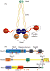
Overall MRN assembly and key domains. (A) MRN can assemble as a heterohexamer and consists of 4 key regions: the processing “head”, formed by the Mre11 dimer and two Rad50 ABC ATPase domains (indicated by dotted circle), the “coil” and “hook” encoded by the region of Rad50 separating the N- and C-terminal ABC ATPase halves, and the Nbs1 “flexible adapter” (indicated by dotted circle) that provides the key link to signaling functions. (B) Schematic representations of the MRN subunits Mre11, Rad50 and Nbs1 showing key domains, colored as in other figures. The approximate location of reported methylation sites are indicated by M and DNA damage inducible phosphorylation sites by P (see text for details). The major sites corresponding to inherited human disorders associated with each gene are indicated by a red triangle, with amino acid substitutions labeled for Mre11 and Rad50 (X is a stop codon) and 657del5 representing the major Nbs1 mutation responsible for >90% of NBS cases.
Figure 3
Figure 3
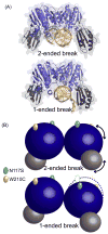
The Mre11 dimer adopts different conformational states at a 2-ended Vs 1-ended DSB. (A) Crystal structures of Mre11 in complex with a 2-ended DSB (top) or a 1-ended DSB replication fork mimic (bottom) revealed Mre11 binds these DNA substrates as symmetric and asymmetric dimers respectively. (B) Cartoon of Mre11 bound to DNA substrates as in (A) with phosphodiesterase and capping domains colored blue and gray, respectively, as in Fig. 1B. Arrows highlight the Mre11 phosphodiesterase and DNA capping domain rotations required to move between 2-ended and 1-ended bound states. ATLD sites that reduce Nbs1 binding (N117S and W210C, green and yellow respectively) are mapped onto the surface and the dotted outline of the symmetric Mre11 dimer is overlaid on the asymmetric dimer to highlight rearrangements between the two states.
Figure 4
Figure 4
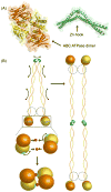
Rad50 states include ATP-dependent association of ABC ATPase domains, and Zn-hook mediated inter-and intramolecular dimers. (A) Structures of the Rad50 ABC ATPase dimer, with N- and C-terminal ABC ATPase halves colored orange and yellow respectively, bound to ATP (in red) and the Zn-hook dimerization domain (green). (B) Cartoon showing several possible Rad50 states. The Rad50 Zn-hook can either intermolecularly dimerize Rad50 within a single MRN complex, which connects the Rad50 ABC ATPase domains present within an M2R2 head (top left), or intramolecularly connect two MRN complexes to form a dumbbell-like structure with M2R2 heads at either end (right). In these cartoons Mre11 is outlined as a dotted circle to show that it can bridge Rad50 molecules in the absence of direct Rad50 dimerization through Zn-hook or ATP-mediated connections. ATP-induced dimerization brings together two Rad50 ABC ATPase domains (bottom left), inducing an ~35º subdomain rotation of the C-terminal subdomain (yellow) with respect to the N-terminal subdomain (orange). ATP is indicated in red. The evolutionarily conserved ABC signature motif is in cyan to facilitate visualization of this rotation.
Figure 5
Figure 5
The Nbs1 flexible adapter. (A) The crystal structure of the Nbs1 N-terminal phosphopeptide binding domain containing the structurally apposed FHA (blue), BRCT1 (yellow) and BRCT2 (red) domains. (B) Cartoon showing interaction states mediated by full length Nbs1. The N-terminal domain mediates interactions with phosphoproteins through binding to phosphothreonine (FHA domain) and/or phosphoserine (BRCT repeat domain) consensus motifs. The extended C-terminus, revealed by SAXS, contains adjacent interaction motifs for Mre11 (dark blue) and ATM (green).
Figure 6
Figure 6
The MRN complex assembles as a flexible sensor, signaler and effector for DSB responses. Model of the MRN complex combining structures of Mre11, Rad50 and Nbs1 domains with known regions of conformational change and/or flexibility (curved arrows) and key interactions (straight arrows) as discussed in the text and figures 3–5. CtIP binding to Nbs1 (shown on the left) induces a conformational change within the phosphopeptide binding core that is suitable to signal to Mre11 through a pull on the flexible tether.

References

    1. Hopfner KP, Putnam CD, Tainer J. DNA double-strand break repair from head to tail. Current Opinion in Structural Biology. 2002;12:115–122. - PubMed
    1. Williams RS, Williams JS, Tainer JA. Mre11-Rad50-Nbs1 is a keystone complex connecting DNA repair machinery, double-strand break signaling, and the chromatin template. Biochem Cell Biol. 2007;85:509–520. - PubMed
    1. Bierne H, Ehrlich SD, Michel B. Deletions at stalled replication forks occur by two different pathways. Embo J. 1997;16:3332–3340. - PMC - PubMed
    1. Michel B, Ehrlich SD, Uzest M. DNA double-strand breaks caused by replication arrest. Embo J. 1997;16:430–438. - PMC - PubMed
    1. Sun H, Treco D, Schultes NP, Szostak JW. Double-strand breaks at an initiation site for meiotic gene conversion. Nature. 1989;338:87–90. - PubMed

Publication types

MeSH terms