Review
. 2011;51(1):13-25.
doi: 10.1016/j.advenzreg.2010.09.008. Epub 2010 Oct 28.

Diphosphoinositol polyphosphates: what are the mechanisms?

Affiliations
Review

Diphosphoinositol polyphosphates: what are the mechanisms?

Stephen B Shears et al. Adv Enzyme Regul. 2011.

Abstract

In countries where adulthood is considered to be attained at age eighteen, 2011 can be the point at which the diphosphoinositol polyphosphates might formally be described as "coming of age", since these molecules were first fully defined in 1993 (Menniti et al., 1993; Stephens et al., 1993b). But from a biological perspective, these polyphosphates cannot quite be considered to have matured into the status of being independently-acting intracellular signals. This review has discussed several of the published proposals for mechanisms by which the diphosphoinositol polyphosphates might act. We have argued that all of these hypotheses need further development.We also still do not know a single molecular mechanism by which a change in the levels of a particular diphosphoinositol polyphosphate can be controlled. Yet, despite all these gaps in our understanding, there is an enduring anticipation that these molecules have great potential in the signaling field. Reflecting our expectations of all teenagers, it should be our earnest hope that in the near future the diphosphoinositol polyphosphates will finally grow up.


Figures
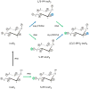
Fig. 1
Fig. 1. Synthesis of diphosphoinositol polyphosphates
These metabolic reactions account for the synthesis of diphosphoinositol polyphosphates in both yeasts and mammalian cells. In the abbreviations of the chemical structures, “Ins” indicates the myo-inositol skeleton. The number of monophosphates around the inositol ring is denoted as a suffix after the ‘P’. The prefixes denote the number of diphosphates (PP). The schematic shows reactions catalyzed by the IP5K (also known as IPK1; black arrow), the IP6Ks (green arrows), and the Vip1/PPIP5Ks (blue arrows). The position of the diphosphate at the 1-position is an arbitrary choice between the two available options, namely, the 1- and 3-positions (see text for details). So as to simplify the figure, the reactions catalyzed by the DIPP phosphatases are not shown.
Fig. 2
Fig. 2. Evidence that diphosphoinositol polyphosphates transphosphorylate proteins in intact cells?
The graphic depicts one particular site on AP3B1 (colored blue) that, in vivo, casein kinase II can mono-phosphorylate, thereby priming it to be transphosphorylated by PP-InsP5 (AP3B1 has other potential phosphorylation sites (Azevedo et al., 2009) that are not illustrated here). Panel a illustrates that Azevedo et al., (Azevedo et al., 2009) heterologously expressed AP3B1 in a kcs1Δ strain of S. cerevisiae. The protein was then extracted and incubated with [32P]PP-InsP5 in vitro (“assay input”). The [32P] (colored red) was transferred from PP-InsP5 to AP3B1 (“assay output”) which can only have occurred if AP3B1 were to have already been mono-phosphorylated by casein kinase II in vivo. Panels b, c depict two alternative explanations for the outcome of separate experiments in which AP3B1 was expressed in either wild-type or vip1Δ S. cerevisiae, which respectively contain either normal or elevated levels of PP-InsP5. AP3B1 was then extracted and incubated with [32P]PP-InsP5 in vitro (“assay input”). Little or no [32P] was transferred from [32P]PP-InsP5 to AP3B1 (“assay output”). There are two explanations for that result. Either (panel b) the AP3B1 was already transphosphorylated (colored black) in vivo, as argued by Azevedo et al., (Azevedo et al., 2009), or (panel c) as we alternately propose, the AP3B1 may not have been monophosphorylated by casein kinase II in vivo. The data do not distinguish between these two possibilities.

Similar articles

Cited by

References

    1. Albert C, Safrany ST, Bembenek ME, Reddy KM, Reddy KK, Falck JR, et al. Biological variability in the structures of diphosphoinositol polyphosphates in Dictyostelium discoideum and mammalian cells. Biochem J. 1997;327:553–560. - PMC - PubMed
    1. Alfieri RR, Petronini PG. Hyperosmotic stress response: comparison with other cellular stresses. Pflugers Arch. 2007;454:173–185. - PubMed
    1. Ali N, Duden R, Bembenek ME, Shears SB. The interaction of coatomer with inositol polyphosphates is conserved in Saccharomyces cerevisiae. Biochem J. 1995;310:279–284. - PMC - PubMed
    1. Azevedo C, Burton A, Ruiz-Mateos E, Marsh M, Saiardi A. Inositol pyrophosphate mediated pyrophosphorylation of AP3B1 regulates HIV-1 Gag release. Proc Natl Acad Sci U S A. 2009;106:21161–21166. - PMC - PubMed
    1. Barker CJ, Illies C, Gaboardi GC, Berggren PO. Inositol pyrophosphates: structure, enzymology and function. Cell Mol Life Sci. 2009;66:3851–3871. - PMC - PubMed

Publication types

Substances

LinkOut - more resources