. 2011 Feb;175(2):133-43.
doi: 10.1016/j.molbiopara.2010.10.007. Epub 2010 Oct 28.

An insertional trap for conditional gene expression in Toxoplasma gondii: identification of TAF250 as an essential gene

Affiliations

An insertional trap for conditional gene expression in Toxoplasma gondii: identification of TAF250 as an essential gene

Lauren Jammallo et al. Mol Biochem Parasitol. 2011 Feb.

Abstract

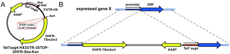
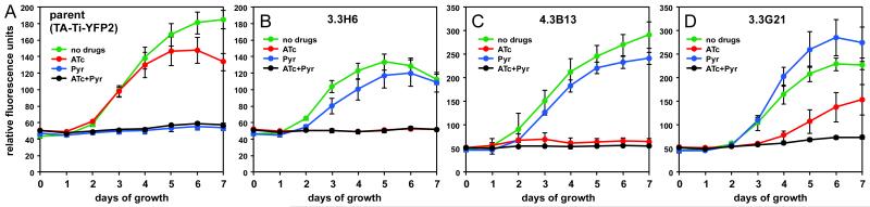
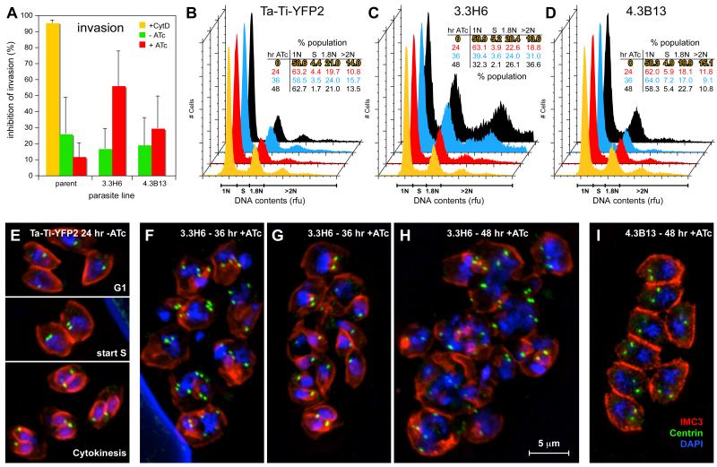
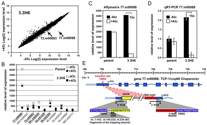
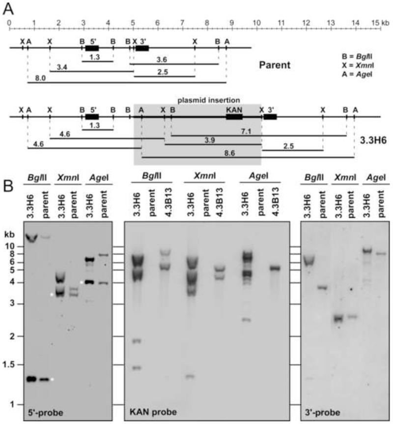
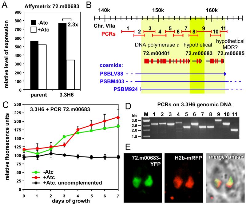
Toxoplasmosis is characterized by fast lytic replication cycles leading to severe tissue lesions. Successful host cell invasion is essential for pathogenesis. The division cycle of Toxoplasma gondii is characterized by an unusual cell cycle progression and a distinct internal budding mechanism. To identify essential genes involved in the lytic cycle we devised an insertional gene trapping strategy using the Tet-transactivator system. In essence, a random, active promoter is displaced with a tetracycline regulatable promoter, which if in an essential gene, will result in a conditionally lethal phenotype upon tetracycline addition. We isolated eight mutants with growth defects, two of which displayed modest invasion defects, one of which had an additional cell cycle defect. The trapped loci were identified using expression microarrays, exploiting the tetracycline dependent expression of the trapped genes. In mutant 3.3H6 we identified TCP-1, a component of the chaperonin protein folding machinery under the control of the Tet promoter. However, this gene was not critical for growth of mutant 3.3H6. Subsequently, we identified a suppressor gene encoding a protein with a hypothetical function by guided cosmid complementation. In mutant 4.3B13, we identified TAF250, an RNA polymerase II complex component, as the trapped, essential gene. Furthermore, by mapping the plasmid insertion boundaries we identified multiple genomic rearrangements, which hint at a potential replication dependent DNA repair mechanism. Furthermore, these rearrangements provide an explanation for inconsistent locus rescue results observed by molecular biological approaches. Taken together, we have added an approach to identify and study essential genes in Toxoplasma.


Figures

Fig. 1
Fig. 1
Schematic outline of insertional trapping with the Tet7sag4 conditional promoter. A. Map of trapping plasmid. The plasmid is 5351 bp long, whereas the ApaI/NdeI restriction site flanked linker is 439 bp long. B. Strategy of mutant generation. The 3′UTR terminator of HXGPRT is removed before transfection by restriction digestion with NdeI and ApaI and purified by gel electrophoresis. To avoid in-fusion insertion in introns, the DHFR ORF is preceded by stop-codons in the three reading frames.
Fig. 2
Fig. 2
Growth kinetics of representative mutants. Growth curves of the parent line (A) and mutants 3.3H6 (B), 4.3B13 (C) and 3.3G21 (D) under various drug pressures. Mutants enter growth arrest when ATc is in the medium, whereas the parent line grows undisturbed. Cytoplasmically expressed tandem YFP was used as a daily read-out in a 384-well plate assay [12]. Error bars denote standard deviation of four replicates.
Fig. 3
Fig. 3
Phenotypic characterization of growth arrest of mutants 3.3H6 and 4.3B13. A. Invasion assays of parent and mutants in absence and presence of ATc induction for 48 hrs. Cytochalasin D (CytD) is used as an efficient invasion blocking control. Assay essentially as in [15]. Error bars denote standard deviation of three replicates. B-D. Flow cytometry analysis of DNA content of the parent line (B) and mutants 3.3H6 (C) and 4.3B13 (D). Profiles after 0, 24, 36 and 48 hrs of ATc induction are shown in yellow, red, blue and black, respectively. Inserted tables display the population development of the various cell cycle stages as a function of ATc induction time. Population definitions are indicated along the x-axis; rfu denotes relative area fluorescence units. E-I. Immunofluorescence assays with α-IMC3 and α-centrin co-stained with DAPI for 3.3H6 and 4.3B13 at various time points with and without ATc induction as indicated.
Fig. 4
Fig. 4
Identification and mapping of the trapped gene in mutant 3.3H6. A. Affymetrix chip expression by expression profiling of mutant 3.3H6 with or without 33 hrs of ATc induction: gene 77.m00088 displays the highest differential expression. B. Overview of the top 10 genes with the highest relative differential expression pattern. The size of differential signal decreases from left to right. Data for parent and mutant 3.3H6 are shown. Underlined, bold gene names were validated by individual cosmid complementation (* 642.m00040 is not annotated as a gene anymore in the latest version of ToxoDB). C. Affymetrix expression profiling detected a 12-fold ATc-dependent conditional expression of gene 77.m00088 in mutant 3.3H6, whereas no variation is detected in the parent. D. By qRT-PCR a 15-fold ATc-dependent conditional expression of gene 77.m00088 is detected in mutant 3.3H6, whereas no variation is detected in the parent. Levels are relative to actin and data were averaged over 5 independent experiments; error bars denote standard error over five replicate experiments with n=3 per experiment. E. Schematic overview of the plasmid insertion breakpoints in gene 77.m00088 on chromosome V identified by PCR and sequencing. At the 5′-end breakpoint an unexpected 303 bp insertion was identified of which corresponds with three non-continuous fragments of the trapping plasmid. See Supplementary Figure S2 for the complete sequence and annotation.
Fig. 5
Fig. 5
Characterization of gene 77.m00088 and the Tet7sag4 insertion in mutant 3.3H6 by Southern blot. A. Schematic of the 77.m00088 locus in wild-type and mutant 3.3H6 genomes. Plasmid insertion represented by grey-shaded box. Restriction sites of BglII, XmnI and AgeI used for the Southern blots are indicated with the expected restriction fragment sizes. Probe localizations are indicated by black boxes. B. Southern blots hybridized with the 5′-probe, the KAN probe and the 3′-probe as indicated. Mutant 4.3B13 is included in the KAN probe hybridization. The 5′-probe cross-reacts with another, unknown fragment in the genome (bands marked with asterisk). The KAN probe demonstrates at least 2, but possibly 3, independent insertions in 3.3H6, whereas 2 insertions are detected in 4.3B13. The AgeI fragment recognized by the 5′probe in the 3.3H6 mutant is approximately 3 kb longer than expected; all other band sizes are as expected.
Fig. 6
Fig. 6
Identification of gene 72.m00683 as a suppressor of mutant 3.3H6. A. Affymetrix expression profiling detected a 2.3-fold ATc-dependent conditional expression of gene 72.m00683 in mutant 3.3H6, whereas no variation is detected in the parent. B. Schematic overview of chromosome VIIa region containing gene 72.m00683. Three cosmids spanning gene 72.m00683 were used to validate its complementation competence, narrowing down the region of interest to the light yellow box. Subsequent PCR amplification of the genomic region marked by the dark yellow box restricted the region to only gene 72.m00683. In red 11 different primer pairs are indicated that were used to map the site of the trapping plasmid insertion. C. Growth curve of mutant 3.3H6 stably complemented with gene 72.m00683 showing growth restoration in the presence of ATc (red line). Un-induced complemented (green line) and uncomplemented with ATc (black line) are included as well. D. PCR products with the 11 primer pairs indicated in panel B show that all amplify the band sizes corresponding with the genomic locus. As such, no trapping plasmid insertion could be detected. E. Expression of a tubulin promoter driven C-terminal YFP fusion of gene 72.m00683 co-transfected with a H2b-mRFP fusion proteins identifies nuclear localization.
Fig. 7
Fig. 7
Identification and validation of the mutated gene in mutant 4.3B13 A. Affymetrix chip expression profiling analysis of mutant 4.3B13. Relative expression data were collected after 33 hrs of ATc induction or un-induced control; gene 64.m00349 is the obvious outlier. B. Growth curves of mutant 4.3B13 growth restoration with cosmid TOXOI78 encoding gene 64.m00349 (red line). Un-induced complemented (green line) and uncomplemented with ATc (black line) are included as well. C. Schematic overview of chromosome III containing gene 64.m00349 and mapping of the plasmid insertion. The insertion was mapped by the 5 PCR primer pairs indicated in red. Primer pair 5 did not amplify from the 4.3B13 mutant and indicated the insertion is within the annotated ORF. Four in-frame ATG start codons are present within the first exon at positions 75,598, 75,640, 76,140, 77,706 and 77,719. The 5′-insertion site contains an unexpected 999 bp sequence consisting of two different fragments of the trapping plasmid as indicated. A short polyC segment of unknown origin separates these two regions. See Supplementary Figure S3 for the complete sequence and annotation. D. PCR amplifications using primer pairs 1-5 indicated in panel C using either parent line (P) and 4.3B13 mutant (M) DNA as template. Primer pair 5 does not amplify from the mutant. E. Neighbor-joining phylogenetic analysis of full length TAF250 sequences from the indicated species. Human TAF250 was used as outgroup. Bootstrap values are indicated at the nodes. Branch lengths indicate the number of nucleotide substitutions per site.

Similar articles

Cited by

References

    1. Montoya JG, Liesenfeld O. Toxoplasmosis. Lancet. 2004;363:1965–1976. - PubMed
    1. Gubbels MJ, White M, Szatanek T. The cell cycle and Toxoplasma gondii cell division: Tightly knit or loosely stitched? Int J Parasitol. 2008;38:1343–1358. - PubMed
    1. Striepen B, Jordan CN, Reiff S, van Dooren GG. Building the perfect parasite: cell division in apicomplexa. PLoS Pathog. 2007;3:e78. - PMC - PubMed
    1. White MW, Radke J, Conde de Felipe M, Lehmann M. Cell cycle control/parasite division. Horizon Scientific Press; Norwich, UK: 2007.
    1. Meissner M, Schluter D, Soldati D. Role of Toxoplasma gondii myosin A in powering parasite gliding and host cell invasion. Science. 2002;298:837–840. - PubMed

Publication types

LinkOut - more resources