. 2011 Feb 3:174:50-63.
doi: 10.1016/j.neuroscience.2010.10.062. Epub 2010 Oct 28.

Complementary synaptic distribution of enzymes responsible for synthesis and inactivation of the endocannabinoid 2-arachidonoylglycerol in the human hippocampus

Affiliations

Complementary synaptic distribution of enzymes responsible for synthesis and inactivation of the endocannabinoid 2-arachidonoylglycerol in the human hippocampus

A Ludányi et al. Neuroscience. .

Erratum in

  • Neuroscience. 2011 Jul 14;186:220

Abstract

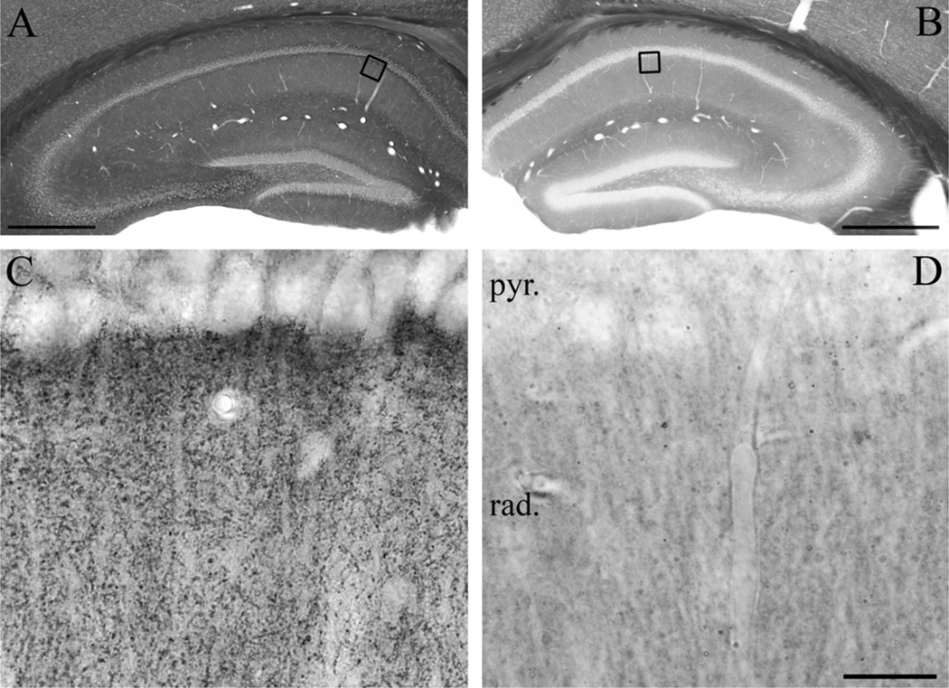
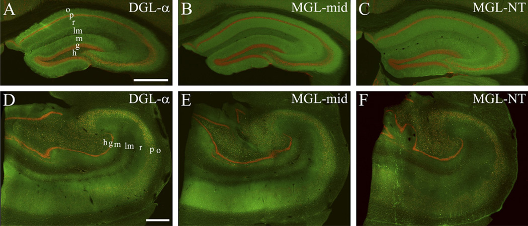
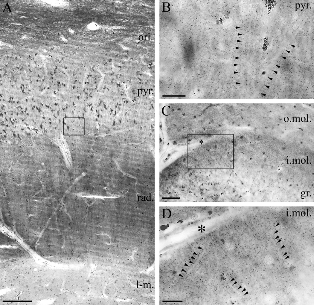
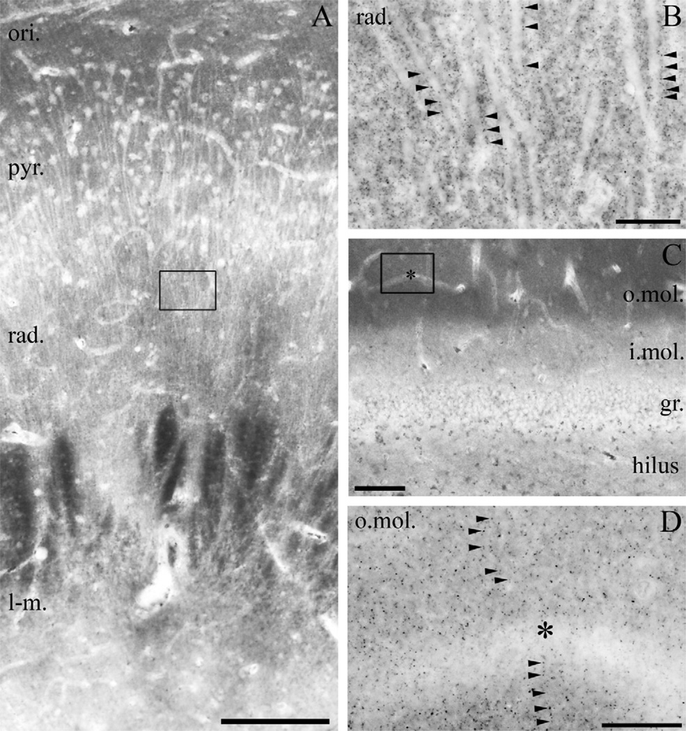
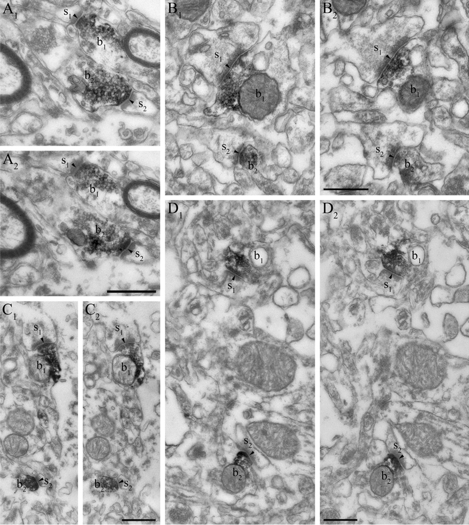
Clinical and experimental evidence demonstrates that endocannabinoids play either beneficial or adverse roles in many neurological and psychiatric disorders. Their medical significance may be best explained by the emerging concept that endocannabinoids are essential modulators of synaptic transmission throughout the central nervous system. However, the precise molecular architecture of the endocannabinoid signaling machinery in the human brain remains elusive. To address this issue, we investigated the synaptic distribution of metabolic enzymes for the most abundant endocannabinoid molecule, 2-arachidonoylglycerol (2-AG), in the postmortem human hippocampus. Immunostaining for diacylglycerol lipase-α (DGL-α), the main synthesizing enzyme of 2-AG, resulted in a laminar pattern corresponding to the termination zones of glutamatergic pathways. The highest density of DGL-α-immunostaining was observed in strata radiatum and oriens of the cornu ammonis and in the inner third of stratum moleculare of the dentate gyrus. At higher magnification, DGL-α-immunopositive puncta were distributed throughout the neuropil outlining the immunonegative main dendrites of pyramidal and granule cells. Electron microscopic analysis revealed that this pattern was due to the accumulation of DGL-α in dendritic spine heads. Similar DGL-α-immunostaining pattern was also found in hippocampi of wild-type, but not of DGL-α knockout mice. Using two independent antibodies developed against monoacylglycerol lipase (MGL), the predominant enzyme inactivating 2-AG, immunostaining also revealed a laminar and punctate staining pattern. However, as observed previously in rodent hippocampus, MGL was enriched in axon terminals instead of postsynaptic structures at the ultrastructural level. Taken together, these findings demonstrate the post- and presynaptic segregation of primary enzymes responsible for synthesis and elimination of 2-AG, respectively, in the human hippocampus. Thus, molecular architecture of the endocannabinoid signaling machinery supports retrograde regulation of synaptic activity, and its similar blueprint in rodents and humans further indicates that 2-AG's physiological role as a negative feed-back signal is an evolutionarily conserved feature of excitatory synapses.


Figures

Fig. 1
Fig. 1
Confirmation of the specificity of the “DGL-α-INT” antibody in DGL-α knockout mice. (A) Immunostaining in wild-type mice using an antibody raised against a 118 residue-long intracellular segment of the DGL-α protein visualizes the layered structure of the hippocampus. The dendritic layers contain high density of DGL-α immunoreactivity, whereas the somatic layers stand out as DGL-α immunonegative. (B) In contrast, immunostaining in the hippocampus of a DGL-α knockout mouse shows no similar immunoreactive profiles, demonstrating antibody specificity. The sections presented in (A) and (B) were incubated in parallel throughout the immunostaining and dehydration procedures to ensure unequivocal comparison. Note the similar intensity of dark tone in the white matter generated by osmification, which confirms identical treatment. Open boxes denote the positions of corresponding insets in the CA1 subfield (C in A, D in B). (C) At higher magnification, immunoreactivity for DGL-α covers the stratum radiatum in wild-type mice. The characteristic punctate staining outlines the major apical trunk of pyramidal cell dendrites, which along with pyramidal cell somata are largely devoid of DGL-α immunoreactivity. (D) The dense DGL-α immunopositive staining in the neuropil is absent in the hippocampus of DGL-α knockout mice. Abbreviations: rad, stratum radiatum; pyr, stratum pyramidale. Scale bars: (A–B) 500 µm; (C–D, shown in D) 20 µm.
Fig. 2
Fig. 2
Comparable topographical distribution of 2-AG metabolizing enzymes in the mouse and human hippocampus. (A–C) Double immunofluorescence staining in the mouse hippocampus shows the laminar organization of DGL-α and MGL, responsible for generation and degradation of the endocannabinoid 2-AG, respectively (green). The cell bodies of neurons are visualized by NeuN staining (red) to highlight the pyramidal and granule cell layers. (D–F) A similar staining pattern is apparent in the human hippocampal formation. Note the laminar organization of the staining pattern for both enzymes indicating the spatial association of 2-AG metabolism with glutamatergic excitatory pathways. “MGL-mid” and “MGL-NT” indicates two distinct primary antibodies raised against different epitopes on the MGL protein. “MGL-mid” gives rise to denser immunostaining in the human hippocampus, whereas “MGL-NT” results in somewhat stronger staining in the mouse hippocampus. Nevertheless, both antibodies reveal comparable overall distribution of MGL in the mouse and human hippocampal formation. Abbreviations: o, stratum oriens; p, stratum pyramidale; r, stratum radiatum; lm, stratum lacunosum-moleculare; m, stratum moleculare; g, stratum granulosum; h, hilus. Scale bars: (A) 0.5 mm also applies to (B–C); (D) 1 mm also applies to (E–F).
Fig. 3
Fig. 3
Light microscopic localization of DGL-α in the human hippocampus. (A) Light micrograph of DGL-α immunoperoxidase reactivity in the CA1 subfield of the cornu ammonis using the “DGL-α-INT” antibody. The highest density of immunostaining is located in the strata oriens (ori.) and radiatum (rad.), whereas staining intensity is somewhat weaker in the strata lacunosum-moleculare (l-m.) and pyramidale (pyr.). Besides the specific DGL-α immunoreactivity, lipofuscin granules in the cell bodies of pyramidal cells are also observed occasionally, but its relation to DGL-α protein was not considered, because these pigment granules are known to accumulate in neuronal somata during aging. (B) At higher magnification, the main apical dendrites of CA1 pyramidal cells at the border of stratum pyramidale and stratum radiatum appear to be DGL-α immunonegative and are contoured by an intense dotted staining in the neuropil (arrowheads). (C) In the dentate gyrus, the highest density of DGL-α immunostaining is visible in the inner third of stratum moleculare (i.mol.), where the mossy cells, a glutamatergic interneuron type of the dentate gyrus terminate. Modest staining is observable in the outer two-thirds of stratum moleculare (o.mol), whereas immunolabelling was weak in stratum granulosum (gr.). (D) At higher magnification, immunonegative dendrites of granule cells are surrounded by widespread punctate DGL-α immunoreactivity (arrowheads) in inner third of the stratum moleculare of dentate gyrus. Open boxes denote the positions of corresponding insets (B in A, D in C). Asterisks in (C–D) depicts the same capillary used as landmark. Scale bars: (A) 200 µm; (B, D) 20 µm; (C) 50 µm.
Fig. 4
Fig. 4
DGL-α is localized in dendritic spines in the mouse and human hippocampus. (A) Immunoperoxidase reaction performed on hippocampal sections derived from wild-type mice reveals that DGL-α is present in numerous dendritic spine heads (s1–s2) receiving asymmetric synapses (arrowheads) in the stratum radiatum of the CA1 subfield. These DGL-α immunopositive spines protrude from DGL-α immunonegative dendritic shafts (d). Other subcellular profiles like excitatory axon terminals (b1–b2) or other dendritic spines (asterisk) are also DGL-α immunonegative, which reflects that this enzyme is either absent or its level is under detection threshold in these compartments. (B) In hippocampal sections derived from DGL-α knockout animals, dendritic spines (asterisks) as well as other subcellular profiles (e.g. dendritic shafts depicted by “d”) are all devoid of DGL-α immunoreactivity confirming the specificity of the antibody for DGL-α. (C1–C2) Serial high-resolution electron micrographs of DGL-α immunoreactivity in the stratum radiatum of the CA1 region in the human hippocampus. The black immunoreaction end-product DAB, representing the localization site of DGL-α, is accumulated in the dendritic spine heads (s1–s2) of CA1 pyramidal neurons. In contrast, DGL-α immunoreactivity levels did not reach detection threshold in presynaptic axon terminals (b1–b2) forming asymmetric synapses (arrowheads) on the spine heads. (D1–D2) Similar situation is found in the inner third of stratum moleculare of the human dentate gyrus. The striking targeting of DGL-α to the postsynaptic side of excitatory synapses (arrowheads) is highlighted by the dense labeling in spine heads (s1–s2), which are derived from DGL-α immunonegative dendritic shafts (d) of granule cells. Other subcellular domains, e.g. axon terminals (b1–b2) are also devoid of DGL-α. Scale bars: (A, B, D2) 500 nm, (D2) also applies to (C1, C2, D1).
Fig. 5
Fig. 5
Light microscopic localization of MGL in the human hippocampus. (A) Light micrograph of immunoperoxidase staining for MGL in the CA1 subfield of the cornu ammonis shows that the highest density of MGL-immunoreactivity using the “MGL-mid” antibody is located in the stratum oriens (ori.), but significant levels of immunostaining is also apparent in strata pyramidale (pyr.), radiatum (rad.) and lacunosum-moleculare (l-m.). (B) At higher magnification, the abundant neuropil staining becomes visible, e.g. in the stratum radiatum of CA1 region (depicted here). The characteristic MGL-immunopositive granules (arrowheads) decorate MGL-immunonegative apical dendrites of pyramidal cells. (C) In the dentate gyrus, MGL-immunostaining was observed predominantly in the outer two thirds of the stratum moleculare (o.mol.), whereas it was weaker in the inner third of stratum moleculare (i. mol.), stratum granulosum (gr.) and in the hilus. Somata of granule cells (similarly to somata of pyramidal cells in A) are hardly or not at all labeled by MGL immunostaining. (D) At higher magnification, the punctate neuropil staining is present throughout the stratum moleculare of the dentate gyrus. MGL-immunonegative dendrites are frequently outlined by the characteristic MGL-containing varicosities often arranged in an array-like manner. Open boxes denote the positions of corresponding insets (B in A, D in C). Asterisks in (C–D) depicts the same capillary used as landmark. Scale bars: (A) 200 µm; (B, D) 20 µm; (C) 100 µm.
Fig. 6
Fig. 6
MGL is located presynaptically on glutamatergic axon terminals in the human hippocampus. (A1–D2) At the electron microscopic level, immunostaining with two independent antibodies raised against different epitopes of the MGL protein reveals the abundance of MGL in axon terminals forming asymmetric, presumably excitatory synapses (arrowheads). The antibody “MGL-mid” is used in (A1–A2) and in (C1–C2), whereas the antibody “MGL-NT” is used in (B1–B2) and in (D1–D2). Series of consecutive ultrathin sections show that the black DAB end product fills the intracellular side of the boutons (b1–b2, all images are two consecutive sections) indicating high levels of MGL enzyme in these axon terminals. Importantly, MGL-immunoreactivity levels did not reach detection threshold in spine heads, in dendritic or in glial processes, and in cell bodies. The illustrated boutons are derived from the stratum radiatum in the CA1 region (b1–b2 in A1–A2 and B1–B2), and from the outer two thirds of stratum moleculare in the dentate gyrus (b1–b2 in C1–C2 and D1–D2). Scale bars: (shown in A2, B2, C2, D2) 500 nm, (also applies to A1, B1, C1, D1, respectively).

Similar articles

Cited by

References

    1. Ahn K, Johnson DS, Mileni M, Beidler D, Long JZ, McKinney MK, Weerapana E, Sadagopan N, Liimatta M, Smith SE, Lazerwith S, Stiff C, Kamtekar S, Bhattacharya K, Zhang Y, Swaney S, Van Becelaere K, Stevens RC, Cravatt BF. Discovery and characterization of a highly selective FAAH inhibitor that reduces inflammatory pain. Chem Biol. 2009;16:411–420. - PMC - PubMed
    1. Ahn K, McKinney MK, Cravatt BF. Enzymatic pathways that regulate endocannabinoid signaling in the nervous system. Chem Rev. 2008;108:1687–1707. - PMC - PubMed
    1. Alexander SP, Kendall DA. The complications of promiscuity: endocannabinoid action and metabolism. Br J Pharmacol. 2007;152:602–623. - PMC - PubMed
    1. Bhaskaran MD, Smith BN. Cannabinoid-mediated inhibition of recurrent excitatory circuitry in the dentate gyrus in a mouse model of temporal lobe epilepsy. PLoS One. 2010;5:e10683. - PMC - PubMed
    1. Bisogno T, Burston JJ, Rai R, Allara M, Saha B, Mahadevan A, Razdan RK, Wiley JL, Di Marzo V. Synthesis and pharmacological activity of a potent inhibitor of the biosynthesis of the endocannabinoid 2-arachidonoylglycerol. Chem Med Chem. 2009;4:946–950. - PubMed

Publication types