Multicenter Study
. 2010 Nov 15;68(10):903-12.
doi: 10.1016/j.biopsych.2010.08.025.

YKL-40: a novel prognostic fluid biomarker for preclinical Alzheimer's disease

Affiliations
Multicenter Study

YKL-40: a novel prognostic fluid biomarker for preclinical Alzheimer's disease

Rebecca Craig-Schapiro et al. Biol Psychiatry. .

Erratum in

  • Biol Psychiatry. 2011 Feb 15;69(4):389

Abstract

Background: Disease-modifying therapies for Alzheimer's disease (AD) would be most effective during the preclinical stage (pathology present, cognition intact) before significant neuronal loss occurs. Therefore, biomarkers that detect AD pathology in its early stages and predict dementia onset and progression will be invaluable for patient care and efficient clinical trial design.

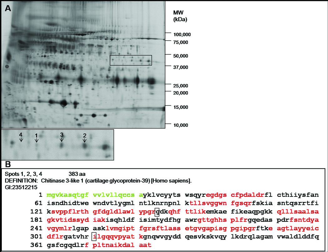
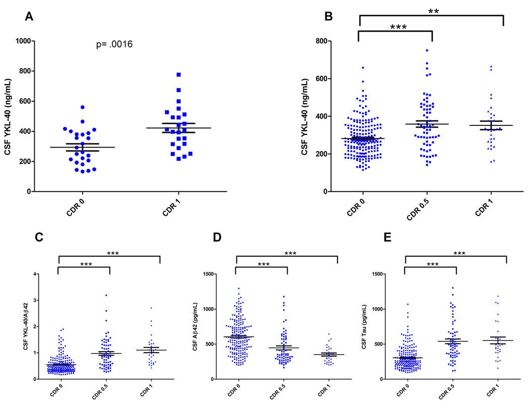
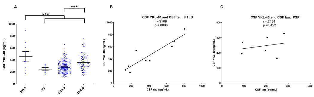
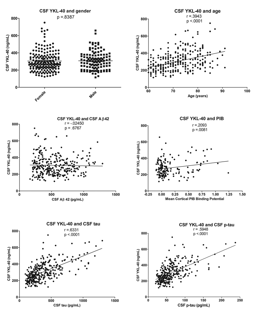
Methods: AD-associated changes in cerebrospinal fluid (CSF) were measured using two-dimensional difference gel electrophoresis and liquid chromatography tandem mass spectrometry. Subsequently, CSF YKL-40 was measured by enzyme-linked immunosorbent assay in the discovery cohort (n = 47), validation cohort (n = 292) with paired plasma samples (n = 237), frontotemporal lobar degeneration (n=9) [corrected], and progressive supranuclear palsy (PSP; n = 6). Immunohistochemistry was performed to identify source(s) of YKL-40 in human AD brain.

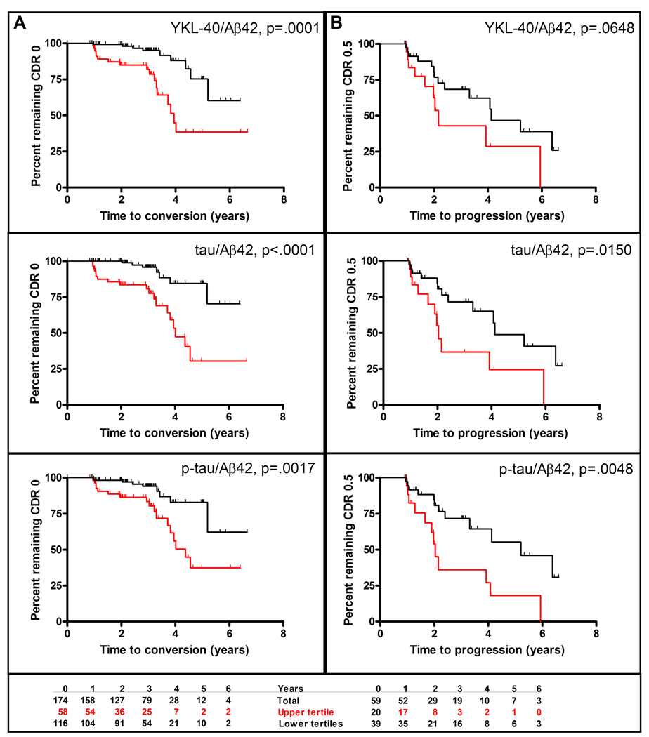
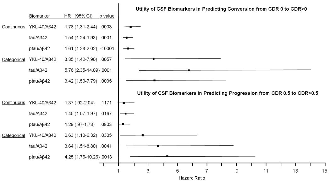
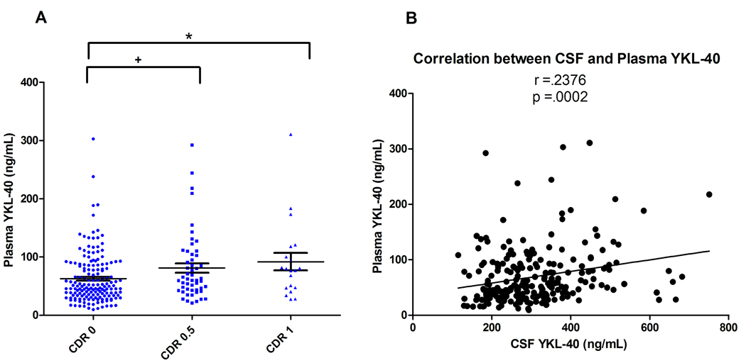
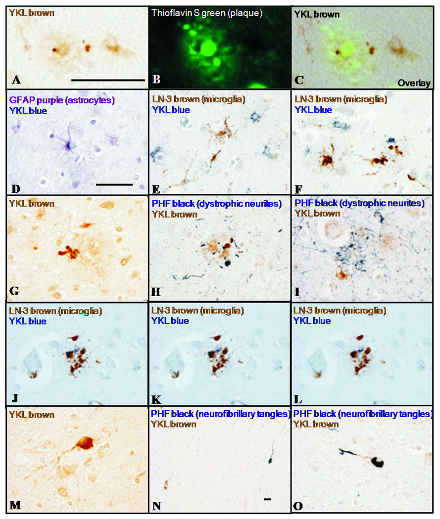
Results: Discovery and validation cohorts, showed higher mean CSF YKL-40 in very mild and mild AD-type dementia (Clinical Dementia Rating [CDR] 0.5 and 1) versus control subjects (CDR 0) and PSP subjects. Importantly, CSF YKL-40/Aβ42 ratio predicted risk of developing cognitive impairment (CDR 0 to CDR > 0 conversion), as well as the best CSF biomarkers identified to date, tau/Aβ42 and p-tau 181/Aβ42. Mean plasma YKL-40 was higher in CDR 0.5 and 1 versus CDR 0, and correlated with CSF levels. YKL-40 immunoreactivity labeled astrocytes near a subset of amyloid plaques, implicating YKL-40 in the neuroinflammatory response to Aβ deposition.

Conclusions: These data demonstrate that YKL-40, a putative indicator of neuroinflammation, is elevated in AD and, together with Aβ42, has potential prognostic utility as a biomarker for preclinical AD.


Figures

Figure 1
Figure 1
(A) A representative 2-D DIGE image of CSF from the discovery cohort. Samples were depleted of six highly abundant proteins, fluorescently labeled, and subjected to isoelectric focusing followed by SDS-PAGE. YKL-40 is more abundant in four spots in the CDR 1 group (labeled 1–4 in the inset, with mean fold changes of 1.41, 1.50, 1.46, 1.32, respectively). The near invisibility of spot 4 in this printed representation illustrates the great sensitivity of 2-D DIGE to detect proteins of low abundance. (B) Sequence coverage of human YKL-40 by mass spectrometry. Indicated in red is the compilation of peptides identified in the four spots. The signal sequence is shown in green, and polymorphisms are indicated by boxes.
Figure 2
Figure 2
Mean YKL-40 is increased in the CSF of CDR 0.5 and CDR 1 subjects. (A) CSF from the discovery cohort (CDR 0, N= 24; CDR 1, N=23) was analyzed for YKL-40 by ELISA (CDR 0= 293.6 +/− 23.9; CDR 1= 422.2 +/− 30.0, ng/mL, mean +/− SEM). CSF YKL-40 was significantly higher in the CDR 1 group as compared to the CDR 0 group (p=.0016, unpaired student’s t-test). (B) CSF from a larger, independent sample set (N=292) was analyzed for YKL-40 by ELISA. Mean CSF YKL-40 was significantly higher in the CDR 0.5 and CDR 1 groups as compared to the CDR 0 group (** p=.004, *** p<.0001; One-way ANOVA with Welch’s correction for unequal variances, Tukey post-hoc Test) (CDR 0= 282.1 +/- 6.7; CDR 0.5= 358.9 +/− 16.9; CDR 1= 351.7 +/− 22.6, ng/mL, mean +/− SEM). (C) Mean CSF YKL-40/Aβ42 was significantly higher in the CDR 0.5 and CDR 1 groups as compared to the CDR 0 group (***p<.0001; One-way ANOVA with Welch’s correction for unequal variances, Tukey post-hoc Test). (D & E) Mean CSF Aβ42 was significantly higher while mean CSF tau was significantly lower in the CDR 0.5 and CDR 1 groups as compared to the CDR 0 group (*** p<.0001; Oneway ANOVA with Welch’s correction for unequal variances, Tukey post-hoc Test). The degree of overlap between clinical groups is comparable for all biomarkers evaluated.
Figure 3
Figure 3
(A) CSF samples from subjects with FTLD (N=9) and PSP (N=6) were analyzed for YKL-40 by ELISA, and levels were compared to those of the validation cohort (CDR 0 and CDR>0 [CDR 0.5&1 combined], N=292). Because the groups differed with respect to mean age at LP (FTLD: 59 yrs, PSP: 66 yrs, CDR 0: 71 yrs, CDR 0.5&1: 75 yrs), analyses were adjusted for age. CSF YKL-40 was significantly higher in the FTLD group as compared to the PSP, CDR 0, and CDR>0 groups (*** p<.0001; ANCOVA, LSD post-hoc Test). While not reaching statistical significance (defined here as α=0.05), CSF YKL-40 levels trended lower in the PSP group as compared to the CDR>0 group. (B–C) CSF YKL-40 and CSF tau values correlated strongly in the FTLD group, but did not correlate in the PSP group.
Figure 4
Figure 4
In the validation cohort, CSF YKL-40 levels do not vary based on gender and are not correlated with CSF Aβ42. However, CSF YKL-40 levels are correlated with age, CSF tau, CSF p-tau181, and mean cortical PIB binding potential.
Figure 5
Figure 5
CSF YKL-40/Aβ42, tau/Aβ42, and p-tau/Aβ42 as predictors of (A) conversion from CDR 0 to CDR>0 and (B) progression from CDR 0.5 to CDR>0.5. Kaplan-Meier estimates of rates of conversion and progression are shown with red curves representing the upper tertile and black curves representing the lower two tertiles. The bottom panel shows for the CSF YKL-40/Aβ42 analyses the number of subjects in the upper and lower tertiles at baseline and at each year of follow-up.
Figure 6
Figure 6
Cox proportional hazards models were used to assess the ability of CSF YKL-40/Aβ42, tau/Aβ42, and ptau/Aβ42 to predict (top) conversion from cognitive normalcy (CDR 0) to cognitive impairment (CDR>0) and (bottom) progression from very mild dementia (CDR 0.5) to mild or moderate dementia (CDR>0.5). Biomarker measures were analyzed as both continuous and categorical variables, and were converted to standard Z-scores to allow comparison of hazard ratios between different biomarkers. In evaluating risk, analyses were adjusted for age and gender. Abbreviations: HR, hazard ratio; CI, confidence interval.
Figure 7
Figure 7
Plasma samples of the validation cohort (N=237) were evaluated for YKL-40 by ELISA. (A) Mean plasma YKL-40 was significantly higher in the CDR 0.5 and CDR 1 groups as compared to the CDR 0 group (+ p=.046, * p=.031; One-way ANOVA, Tukey post-hoc Test) (CDR 0= 62.5 +/− 3.4; CDR 0.5= 81.1 +/− 8.0; CDR 1= 91.9 +/− 15.0, ng/mL, mean +/− SEM). (B) CSF and plasma YKL-40 levels are significantly correlated (r =.2376, p=.0002).
Figure 8
Figure 8
In AD neocortex, YKL-40 immunoreactivity is observed in the vicinity of thioflavin S-positive fibrillar amyloid plaques (A,B,C). YKL-40 immunoreactivity is present within a subset of GFAP-positive astrocytes (D) and not in LN-3-positive microglia (E,F). YKL-40 is also observed in cell processes associated with plaques (G); these processes lack reactivity for dystrophic neurite marker PHF-1 (H,I) and microglial marker LN-3 (J,K,L representing adjacent focal planes), and may represent astrocytic processes. YKL-40 immunoreactivity is also observed in occasional neurons in the superficial white matter (M,N,O), some of which contain neurofibrillary tangles (evidenced by PHF-1 staining, N,O). These neurons may represent cells of multiform layer VI or ‘interstitial neurons’ of the white matter. Scale bars = 50 µm; scale bar in A applies to A–C; scale bar in D applies to D–O, with the exception of N.

References

    1. Davies L, Wolska B, Hilbich C, Multhaup G, Martins R, Simms G, et al. A4 amyloid protein deposition and the diagnosis of Alzheimer's disease: prevalence in aged brains determined by immunocytochemistry compared with conventional neuropathologic techniques. Neurology. 1988;38:1688–1693. - PubMed
    1. Morris J, Price J. Pathologic correlates of nondemented aging, mild cognitive impairment, and early stage Alzheimer's disease. J Mol Neurosci. 2001;17:101–118. - PubMed
    1. Hulette CM, Welsh-Bohmer KA, Murray MG, Saunders AM, Mash DC, McIntyre LM. Neuropathological and neuropsychological changes in "normal" aging: Evidence for preclinical Alzheimer disease in cognitively normal individuals. J Neuropathol Exp Neurol. 1998;57:1168–1174. - PubMed
    1. Craig-Schapiro R, Fagan AM, Holtzman DM. Biomarkers of Alzheimer's disease. Neurobiol Dis. 2009;35:128–140. - PMC - PubMed
    1. Perrin RJ, Fagan AM, Holtzman DM. Multimodal techniques for diagnosis and prognosis of Alzheimer's disease. Nature. 2009;461:916–922. - PMC - PubMed

Publication types

MeSH terms