. 2011 Jan 28;300(2):180-8.
doi: 10.1016/j.canlet.2010.10.004. Epub 2010 Oct 29.

Small molecule antagonists for CXCR2 and CXCR1 inhibit human colon cancer liver metastases

Affiliations

Small molecule antagonists for CXCR2 and CXCR1 inhibit human colon cancer liver metastases

Michelle L Varney et al. Cancer Lett. .

Abstract

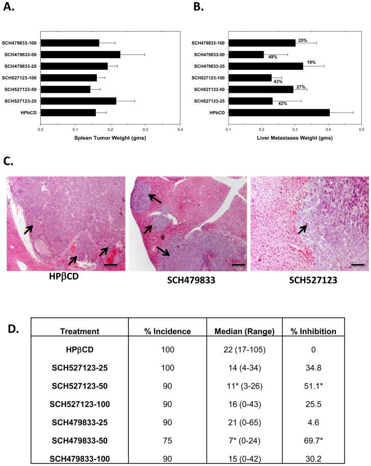
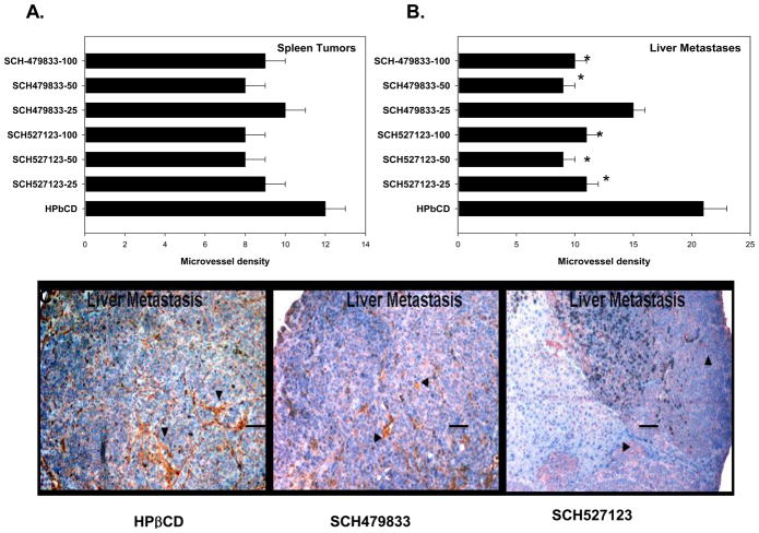
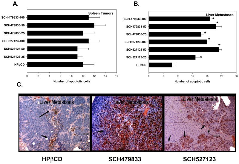
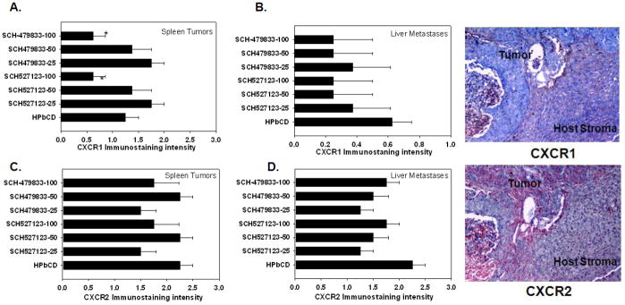
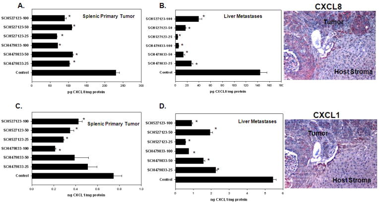
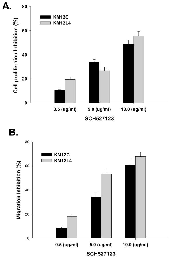
CXCR1 and CXCR2 are G-protein coupled receptors, that have been shown to play important role in tumor growth and metastasis, and are prime targets for the development of novel therapeutics. Here, we report that targeting CXCR2 and CXCR1 activity using orally active small molecule antagonist (SCH-527123, SCH-479833) inhibits human colon cancer liver metastasis mediated by decreased neovascularization and enhanced malignant cell apoptosis. There were no differences in primary tumor growth. These studies demonstrate the important role of CXCR2/1 in colon cancer metastasis and that inhibition of CXCR2 and CXCR1, small molecule antagonists provides a novel therapeutic strategy.


Conflict of interest statement

Conflicts of Interest Statement

None Declared

Figures

Figure 1
Figure 1. SCH-527123 and SCH-479833 inhibited colon cancer liver metastases
A. Primary spleen tumor weights of mice treated with SCH-527123 or SCH-479833. Wet tumor-bearing spleens were weighed and normalized with spleens from normal mice. The values are average normalized weight in grams (gms) ± standard error of mean (SEM). B. Liver metastases in SCH-527123 or SCH-479833 treated animals. The liver metastatic burden was analyzed as described in materials and methods. The values are mean normalized liver metastatic weight ± SEM. C. A representative tumor section showing metastatic nodules in livers of control and antagonist treated mice. Scale bar represents 0.1 mm. D. The incidence of liver metastases. Livers were examined for metastases and the numbers of nodules were counted. The values are the median number of nodules per mouse with the range indicated. The median number of nodules in antagonist treated animals was compared to the control treated group and percent inhibition was calculated. * p<0.05, significantly different from control group.
Figure 2
Figure 2. SCH-527123 and SCH-479833 treatment decreased neovascularization in liver metastases
A. Primary splenic tumors and liver metastatic lesions were immunostained with anti-CD31 antibody and the numbers of microvessels were quantitated. The values are number of microvessel in primary splenic tumors (A) and liver metastases (B). C. A representative liver metastatic nodule showing anti-CD31 staining in control and antagonist SCH527123 (50 MPK) and SCH-479833 (50 MPK) treated mice. * p<0.05, significantly different from control group. Scale bar represents 0.01 mm.
Figure 3
Figure 3. CXCR1 and CXCR2 antagonists treatment increased malignant cell apoptosis
A. Primary tumor and liver metastatic sections were stained for apoptosis using TUNEL and the numbers of apoptotic cells were quantitated. The values are number of apoptotic cells in primary splenic tumors (A) and liver metastases (B). C. A representative liver metastatic section from control and receptor antagonist SCH527123 (50 MPK) and SCH-479833 (50 MPK) treated mice. * p<0.05, significantly different from control group. Scale bar represents 0.01 mm.
Figure 4
Figure 4. Human CXCR1 and CXCR2 expression in splenic lesions and liver metastases
A. CXCR1 expression in splenic lesions. CXCR1 expression was evaluated by immunostaining in primary splenic lesions (A) and liver metastases (B). CXCR2 expression in primary splenic tumors (C) and liver metastatic lesions (D) were analyzed using IHC. The values are mean immunostaining intensity ± SEM. A representative photomicrograph to demonstrate human CXCR1 and CXCR2 immunostaining in SCH527123-100 MPK treated liver metastases to demonstrate expression of receptors in tumor and host stromal compartment. No immunostaining of human CXCR1 and CXCR2 was observed in murine stromal tissue.
Figure 5
Figure 5. Human CXCL8 and CXCL1 expression in splenic and liver lysates
A. CXCL8 expression in splenic lysates. Treatment with receptor antagonists decreased CXCL8 protein levels in spleens. B. CXCL8 expression in liver lysates. CXCL8 levels in liver lysates were significantly decreased in all antagonist treated groups. C. CXCL1 expression in splenic lysates. CXCL1 expression was slightly decreased in splenic lysates after treatment with CXCR2/1 antagonists. D. CXCL1 expression in liver lysates. Treatment with CXCR2/1 receptor antagonists significantly decreased CXCL1 expression in liver lysates. A representative photomicrograph to demonstrate human CXCL1 and CXCL8 immunostaining in SCH527123-100 MPK treated liver metastases to demonstrate expression of receptors in tumor and host stromal compartment. No immunostaining of human CXCL1 and CXCL2 was observed in murine stromal tissue.
Figure 6
Figure 6. Inhibition of cell proliferation and motility in CXCR2/1 antagonist treated cells
A. KM12C and KM12L4 cells (1000 cells/well) in a 96 well plate were cultured in medium with different concentrations of SCH-527123. Cellular proliferation was determined at 72h by MTT assay. The values are mean percent inhibition of proliferation ± SEM. This is a representative of three experiments done in triplicate. B. Cells were plated on non-coated membranes for motility assay, and incubated overnight. Serum free medium containing different concentrations of SCH-527123 was added to the lower chamber. The cells that did not migrate through the Matrigel and/or pores in the membrane were removed, and cells on the other side of the membrane were stained and photographed at 200× magnification. Cells were counted in ten random fields (200×) and expressed as the average number of cells per field of view. The values are number of migrated cells ± SEM.

Similar articles

Cited by

References

    1. Jemal A, Siegel R, Xu J, Ward E. Cancer Statistics. CA Cancer J Clin. 2010:2010. - PubMed
    1. Li A, Varney ML, Singh RK. Expression of interleukin 8 and its receptors in human colon carcinoma cells with different metastatic potentials. Clin Cancer Res. 2001;7:3298–3304. - PubMed
    1. Ruffini PA, Morandi P, Cabioglu N, Altundag K, Cristofanilli M. Manipulating the chemokine-chemokine receptor network to treat cancer. Cancer. 2007;109:2392–2404. - PubMed
    1. Matsushima K, Oppenheim JJ. Interleukin 8 and MCAF: novel inflammatory cytokines inducible by IL 1 and TNF. Cytokine. 1989;1:2–13. - PubMed
    1. Rossi D, Zlotnik A. The biology of chemokines and their receptors. Annu Rev Immunol. 2000;18:217–242. - PubMed

Publication types

MeSH terms

Substances