

The social brain in adolescence: evidence from functional magnetic resonance imaging and behavioural studies
- PMID: 21036192
- PMCID: PMC4538788
- DOI: 10.1016/j.neubiorev.2010.10.011
The social brain in adolescence: evidence from functional magnetic resonance imaging and behavioural studies
Abstract
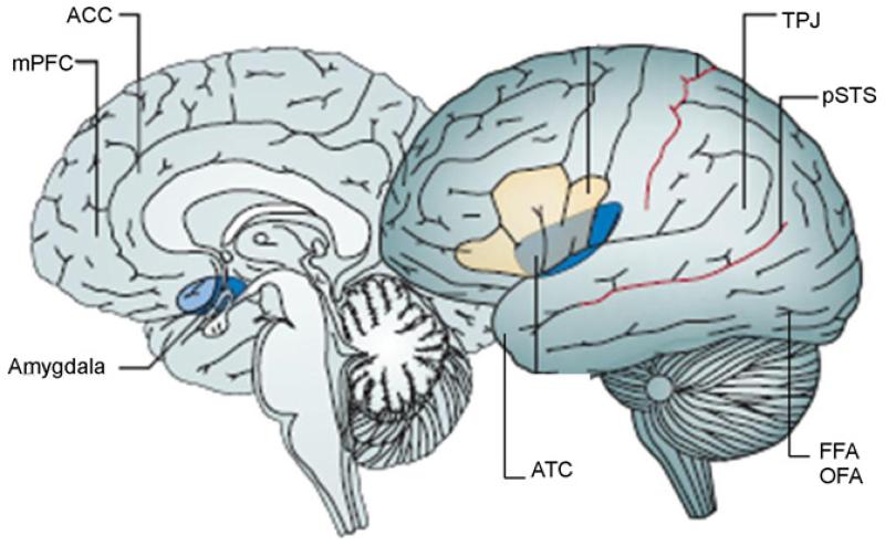
Social cognition is the collection of cognitive processes required to understand and interact with others. The term 'social brain' refers to the network of brain regions that underlies these processes. Recent evidence suggests that a number of social cognitive functions continue to develop during adolescence, resulting in age differences in tasks that assess cognitive domains including face processing, mental state inference and responding to peer influence and social evaluation. Concurrently, functional and structural magnetic resonance imaging (MRI) studies show differences between adolescent and adult groups within parts of the social brain. Understanding the relationship between these neural and behavioural observations is a challenge. This review discusses current research findings on adolescent social cognitive development and its functional MRI correlates, then integrates and interprets these findings in the context of hypothesised developmental neurocognitive and neurophysiological mechanisms.
Crown Copyright © 2010. Published by Elsevier Ltd. All rights reserved.
Figures
References
-
- Abraham A, Werning M, Rakoczy H, von Cramon DY, Schubotz RI. Minds, persons, and space: an fMRI investigation into the relational complexity of higher-order intentionality. Consciousness and Cognition. 2008;17(2):438–450. - PubMed
-
- Amodio DM, Frith CD. Meeting of minds: the medial frontal cortex and social cognition. Nature Reviews Neuroscience. 2006;7(4):268–277. - PubMed
-
- Ashburner J, Friston KJ. Voxel-based morphometry—the methods. NeuroImage. 2000;11(6):805–821. - PubMed
-
- Attwell D, Iadecola C. The neural basis of functional brain imaging signals. Trends in Neurosciences. 2002;25(12):621–625. - PubMed
Publication types
MeSH terms
Grants and funding
LinkOut - more resources
Full Text Sources
Medical
