

Detection of granularity in dermoscopy images of malignant melanoma using color and texture features
- PMID: 21036538
- PMCID: PMC3159567
- DOI: 10.1016/j.compmedimag.2010.09.005
Detection of granularity in dermoscopy images of malignant melanoma using color and texture features
Abstract
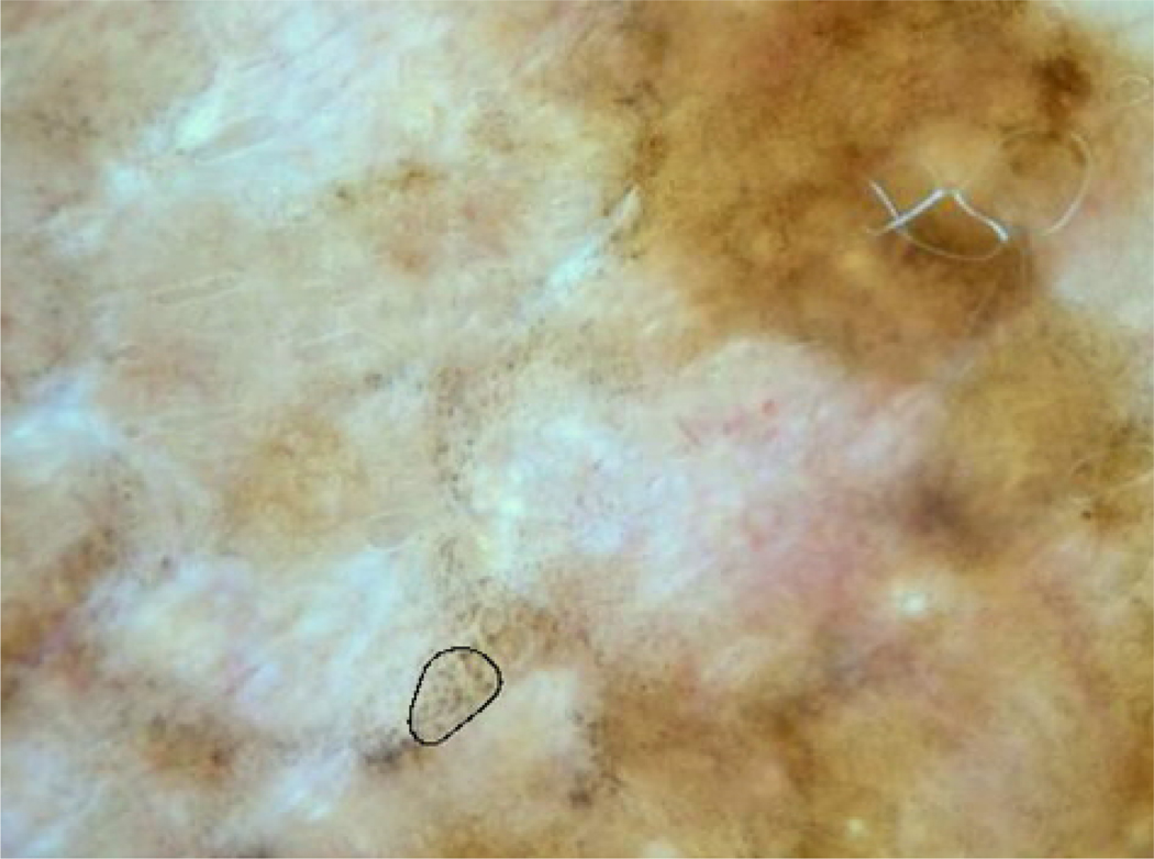
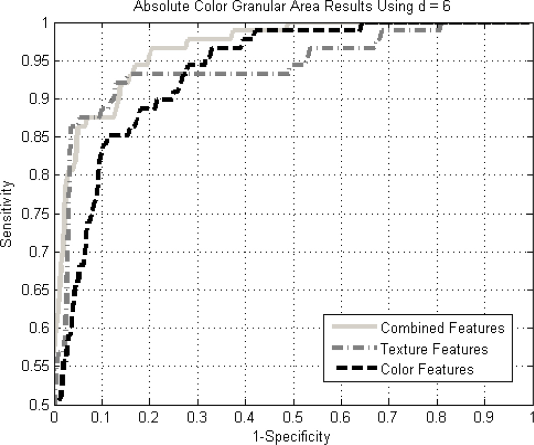
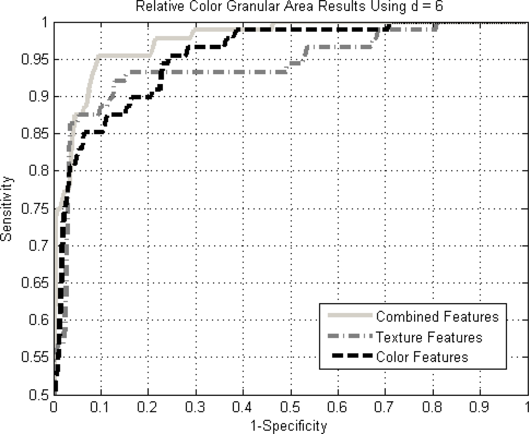
Granularity, also called peppering and multiple blue-grey dots, is defined as an accumulation of tiny, blue-grey granules in dermoscopy images. Granularity is most closely associated with a diagnosis of malignant melanoma. This study analyzes areas of granularity with color and texture measures to discriminate granularity in melanoma from similar areas in non-melanoma skin lesions. The granular areas in dermoscopy images of 74 melanomas and 14 melanomas in situ were identified and manually selected. For 200 non-melanoma dermoscopy images, those areas which most closely resembled granularity in color and texture were similarly selected. Ten texture and twenty-two color measures were studied. The texture measures consisted of the average and range of energy, inertia, correlation, inverse difference, and entropy. The color measures consisted of absolute and relative RGB averages, absolute and relative RGB chromaticity averages, absolute and relative G/B averages, CIE X, Y, Z, X/Y, X/Z and Y/Z averages, R variance, and luminance. These measures were calculated for each granular area of the melanomas and the comparable areas in the non-melanoma images. Receiver operating characteristic (ROC) curve analysis showed that the best separation of melanoma images from non-melanoma images by granular area features was obtained with a combination of color and texture measures. Comparison of ROC results showed greater separation of melanoma from benign lesions using relative color than using absolute color. Statistical analysis showed that the four most significant measures of granularity in melanoma are two color measures and two texture measures averaged over the spots: relative blue, relative green, texture correlation, and texture energy range. The best feature set, utilizing texture and relative color measures, achieved an accuracy of 96.4% based on area under the receiver operating characteristic curve.
Copyright © 2010 Elsevier Ltd. All rights reserved.
Figures
References
-
- Argenziano G, Soyer HP, Chimenti S, Talamini R, Corona R, Sera F, et al. Dermoscopy of pigmented skin lesions: Results of a consensus meeting via the Internet. J Am Acad Dermatol. 2003;48:679–693. - PubMed
-
- Braun RP, Gaide O, Oliviero M, Kopf AW, French LE, Saurat JH, Rabinovitz HS. The significance of multiple blue-grey dots (granularity) for the dermoscopic diagnosis of melanoma. Br J Dermatol. 2007;157:907. - PubMed
-
- Zalaudek I, Argenziano G, Ferrara G, Soyer HP, Corona R, Sera F, et al. Clinically equivocal melanocytic skin lesions with features of regression: a dermoscopic-pathological study. British Journal of Dermatology. 2004;150:64. - PubMed
-
- Massi D, De Giorgi V, Carli P, Santucci M. Diagnostic significance of the blue hue in dermoscopy of melanocytic lesions: a dermoscopic-pathological study. Am J Dermatopathol. 2001;23:463. - PubMed
-
- Benvenuto-Andrade C, Dusza SW, Agero AL, Scope A, Rajadhyaksha M, Halpern AC, Marghoob AA. Differences between polarized light dermoscopy and immersion contact dermoscopy for the evaluation of skin lesions. Arch Dermatol. 2007;143:329. - PubMed
Publication types
MeSH terms
Grants and funding
LinkOut - more resources
Full Text Sources
Medical
