

Chemical methods to detect S-nitrosation
- PMID: 21036657
- PMCID: PMC3033964
- DOI: 10.1016/j.cbpa.2010.10.006
Chemical methods to detect S-nitrosation
Abstract
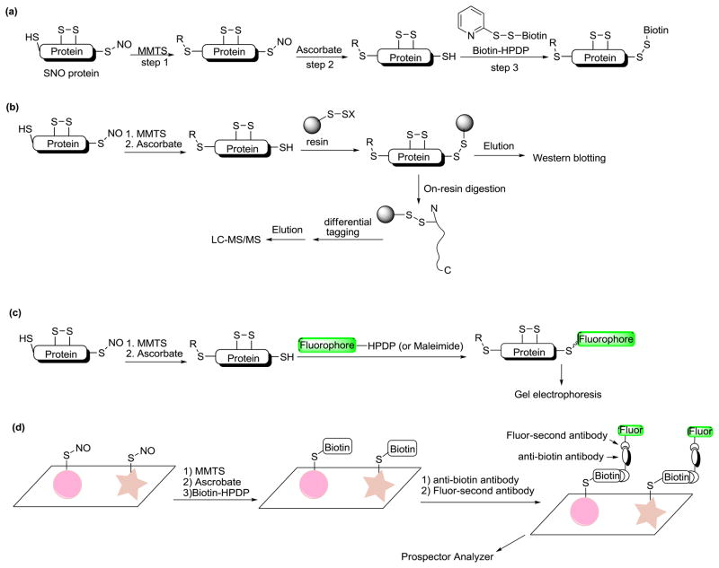
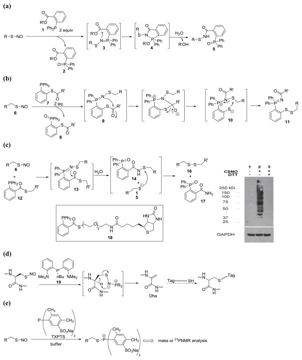
Nitric oxide (NO) is a cell-signaling molecule involved in a number of physiological and pathophysiological processes. Modification of cysteine residues by NO (or NO metabolites), that is S-nitrosation, changes the function of a broad spectrum of proteins. This reaction represents an important post-translational modification that transduces NO-dependent signals. However, the detection and quantification of S-nitrosation in biological samples remain a challenge mainly because of the lability of S-nitrosation products: S-nitrosothiols (SNO). In this review we summarize recent developments of the methods to detect S-nitrosation. Our focus is on the methods which can be used to directly conjugate the site(s) of S-nitrosation.
Copyright © 2010 Elsevier Ltd. All rights reserved.
Figures

Similar articles
-
Identification of novel S-nitrosation sites in soluble guanylyl cyclase, the nitric oxide receptor.J Proteomics. 2016 Apr 14;138:40-7. doi: 10.1016/j.jprot.2016.02.009. Epub 2016 Feb 18. J Proteomics. 2016. PMID: 26917471 Free PMC article.
-
Identification of Protein Targets of S-Nitroso-Coenzyme A-Mediated S-Nitrosation Using Chemoproteomics.ACS Chem Biol. 2024 Jan 19;19(1):193-207. doi: 10.1021/acschembio.3c00654. Epub 2023 Dec 30. ACS Chem Biol. 2024. PMID: 38159293 Free PMC article.
-
Chemical methods for the direct detection and labeling of S-nitrosothiols.Antioxid Redox Signal. 2012 Oct 1;17(7):981-91. doi: 10.1089/ars.2012.4570. Epub 2012 Mar 23. Antioxid Redox Signal. 2012. PMID: 22356122 Free PMC article. Review.
-
Promotion of S-nitrosation of cysteine by a {Co(NO)2}10 complex.Chem Commun (Camb). 2023 Aug 8;59(64):9774-9777. doi: 10.1039/d3cc02784h. Chem Commun (Camb). 2023. PMID: 37486167
-
The chemical biology of S-nitrosothiols.Antioxid Redox Signal. 2012 Oct 1;17(7):969-80. doi: 10.1089/ars.2012.4590. Epub 2012 Jun 7. Antioxid Redox Signal. 2012. PMID: 22468855 Free PMC article. Review.
Cited by
-
Protein S-nitrosylation: specificity and identification strategies in plants.Front Chem. 2015 Jan 7;2:114. doi: 10.3389/fchem.2014.00114. eCollection 2014. Front Chem. 2015. PMID: 25750911 Free PMC article. Review.
-
Methylsulfonyl benzothiazole (MSBT): a selective protein thiol blocking reagent.Org Lett. 2012 Jul 6;14(13):3396-9. doi: 10.1021/ol301370s. Epub 2012 Jun 8. Org Lett. 2012. PMID: 22681565 Free PMC article.
-
(De)Activation (Ir)Reversibly or Degradation: Dynamics of Post-Translational Protein Modifications in Plants.Life (Basel). 2022 Feb 21;12(2):324. doi: 10.3390/life12020324. Life (Basel). 2022. PMID: 35207610 Free PMC article. Review.
-
Specific Reactions of RSNO, HSNO, and HNO and Their Applications in the Design of Fluorescent Probes.Chemistry. 2020 Sep 10;26(51):11673-11683. doi: 10.1002/chem.202001885. Epub 2020 Jul 20. Chemistry. 2020. PMID: 32433809 Free PMC article. Review.
-
Phosphine Mediated Conjugation of S-Nitrosothiols and Aldehydes.Tetrahedron Lett. 2015 May 20;56(21):2741-2743. doi: 10.1016/j.tetlet.2015.04.017. Tetrahedron Lett. 2015. PMID: 26089576 Free PMC article.
References
-
- Gladwin MT, Wang X, Hogg N. Methodological vexation about thiol oxidation versus S-nitrosation-A commentary on “An ascorbate-dependent artifact that interferes with the interpretation of the biotin-switch assay”. Free Radic Biol Med. 561;2006:41–557. The authors point out possible problems of the biotin switch method and suggest that careful control experiments be necessary when biotin switch is used to analyze protein S-nitrosation. - PubMed
-
- Giustarini D, Milzani A, Dale-Donne I, Rossi R. Detection of s-nitrosothiol in biological fluids: a comparison among the most widely applied methodologies. J Chromatogr B Analyt Technol biomed Life Sci. 2007;851:124–139. - PubMed
-
- MacArthur PH, Shiva S, Galdwin MT. Measurement of circulating nitrite and S-nitrosothiols by reductive chemiluminescence. J Chromatogr B Analyt Technol biomed Life Sci. 2007;851:93–105. - PubMed
Publication types
MeSH terms
Substances
Grants and funding
LinkOut - more resources
Full Text Sources
