. 2011 Jan;39(Database issue):D392-401.
doi: 10.1093/nar/gkq1021. Epub 2010 Oct 29.

The RCSB Protein Data Bank: redesigned web site and web services

Affiliations

The RCSB Protein Data Bank: redesigned web site and web services

Peter W Rose et al. Nucleic Acids Res. 2011 Jan.

Abstract

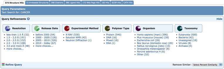
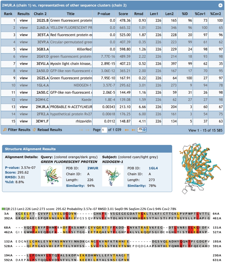
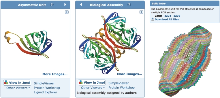
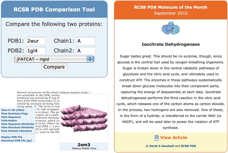
The RCSB Protein Data Bank (RCSB PDB) web site (http://www.pdb.org) has been redesigned to increase usability and to cater to a larger and more diverse user base. This article describes key enhancements and new features that fall into the following categories: (i) query and analysis tools for chemical structure searching, query refinement, tabulation and export of query results; (ii) web site customization and new structure alerts; (iii) pair-wise and representative protein structure alignments; (iv) visualization of large assemblies; (v) integration of structural data with the open access literature and binding affinity data; and (vi) web services and web widgets to facilitate integration of PDB data and tools with other resources. These improvements enable a range of new possibilities to analyze and understand structure data. The next generation of the RCSB PDB web site, as described here, provides a rich resource for research and education.


Figures

Figure 1.
Figure 1.
The Chemical Component Search interface supports four search types. The query structure is shown on the left and an example structure that matches the query is shown on the right. The exact, substructure and superstructure search match the specified stereochemistry, if it is specified in the query. The similarity search ignores stereochemistry.
Figure 2.
Figure 2.
The query-refinement-user interface on the query results page displays the distribution of search hits by various categories. The number of hits for each subcategory is displayed in parenthesis and links to the corresponding subset of hits.
Figure 3.
Figure 3.
Structural alignments. Top: list of representative protein chains that are structurally similar to green fluorescent protein PDB ID 2WUR, chain A (2WUR.A) calculated by jFATCAT. The list is sorted by P-value: significance of alignment; Score: raw alignment; Cα RMSD: the deviation of the aligned residues; Len1: length of the query chain; Len2: length of subject chain; %ID: percent sequence identity; %Cov1: coverage or % of residues in the query chain that are aligned, %Cov2: coverage or % of residues in the subject chain that are aligned. Middle: structural alignment results for green fluorescent protein 2WUR.A (orange) with nidogen-1 1GL4.A (cyan). Unaligned residues are rendered in grey. Bottom: the sequence alignment shows structurally aligned residues highlighted: identical residues (red), similar residues (yellow), other aligned residues (grey) and unaligned residues (white).
Figure 4.
Figure 4.
Structure visualization. Left: the asymmetric unit of streptavidin in PDB entry 1STP. The 3D viewers listed below the image will display the asymmetric unit. The arrow toggles between the asymmetric unit and the biological assembly. Middle: the biological assembly of streptavidin based on oligomeric state assigned by the author. The 3D viewers listed below the image will display the biological assembly. Right: a very large biological assembly, the rat liver vault protein, visualized in the Jmol viewer. The structure is the composite of the three PDB entries 2ZUO, 2ZV4 and 2ZV5 with symmetry transformation (−x, y, −z) applied to generate the complete biological assembly. To accommodate the large size, only the Cα positions are used to render the structure.
Figure 5.
Figure 5.
Several widgets are available for embedding PDB resources into third-party web sites. Top left: the comparison tool that may be used to align sequences or structures, shown here in use for a structural alignment of green fluorescent protein and nidogen-1. Bottom left: a PDB ID in a sample document (excerpt from http://www.pdb.org/pdb/static.do?p=education_discussion/molecule_of_the_month/pdb109_1.html) is marked up with the menu tag that displays a menu to view and download information from the RCSB PDB site. Also shown is an example of the image tag for PDB ID 2OM3. Right: the Molecule of the Month widget that presents the first paragraph of the current Molecule of the Month, and links to the full article.

Similar articles

Cited by

References

    1. Berman HM, Westbrook J, Feng Z, Gilliland G, Bhat TN, Weissig H, Shindyalov IN, Bourne PE. The Protein Data Bank. Nucleic Acids Res. 2000;28:235–242. - PMC - PubMed
    1. Berman H, Henrick K, Nakamura H. Announcing the worldwide Protein Data Bank. Nat. Struct. Biol. 2003;10:980. - PubMed
    1. Velankar S, Best C, Beuth B, Boutselakis CH, Cobley N, Sousa Da Silva AW, Dimitropoulos D, Golovin A, Hirshberg M, John M, et al. PDBe: Protein Data Bank in Europe. Nucleic Acids Res. 2010;38:D308–D317. - PMC - PubMed
    1. Dutta S, Burkhardt K, Young J, Swaminathan GJ, Matsuura T, Henrick K, Nakamura H, Berman HM. Data deposition and annotation at the Worldwide Protein Data Bank. Mol. Biotechnol. 2009;42:1–13. - PubMed
    1. Deshpande N, Addess KJ, Bluhm WF, Merino-Ott JC, Townsend-Merino W, Zhang Q, Knezevich C, Xie L, Chen L, Feng Z, et al. The RCSB Protein Data Bank: a redesigned query system and relational database based on the mmCIF schema. Nucleic Acids Res. 2005;33:D233–D237. - PMC - PubMed

Publication types