. 2011 Mar;39(4):1473-84.
doi: 10.1093/nar/gkq982. Epub 2010 Oct 29.

A discontinuous DNA glycosylase domain in a family of enzymes that excise 5-methylcytosine

Affiliations

A discontinuous DNA glycosylase domain in a family of enzymes that excise 5-methylcytosine

María Isabel Ponferrada-Marín et al. Nucleic Acids Res. 2011 Mar.

Abstract

DNA cytosine methylation (5-meC) is a widespread epigenetic mark associated to gene silencing. In plants, DEMETER-LIKE (DML) proteins typified by Arabidopsis REPRESSOR OF SILENCING 1 (ROS1) initiate active DNA demethylation by catalyzing 5-meC excision. DML proteins belong to the HhH-GPD superfamily, the largest and most functionally diverse group of DNA glycosylases, but the molecular properties that underlie their capacity to specifically recognize and excise 5-meC are largely unknown. We have found that sequence similarity to HhH-GPD enzymes in DML proteins is actually distributed over two non-contiguous segments connected by a predicted disordered region. We used homology-based modeling to locate candidate residues important for ROS1 function in both segments, and tested our predictions by site-specific mutagenesis. We found that amino acids T606 and D611 are essential for ROS1 DNA glycosylase activity, whereas mutations in either of two aromatic residues (F589 and Y1028) reverse the characteristic ROS1 preference for 5-meC over T. We also found evidence suggesting that ROS1 uses Q607 to flip out 5-meC, while the contiguous N608 residue contributes to sequence-context specificity. In addition to providing novel insights into the molecular basis of 5-meC excision, our results reveal that ROS1 and its DML homologs possess a discontinuous catalytic domain that is unprecedented among known DNA glycosylases.


Figures

Figure 1.
Figure 1.
An unusual sequence insertion is present in the DNA glycosylase domain of members of the DML family. (A) Schematic diagram showing ROS1 regions conserved among DML proteins. (B) Multiple sequence alignment of DML proteins and several HhH-GPD superfamily members. Listed above the primary sequence are indicated secondary structure assignments from the ROS1 model prediction shown in (C), colored according to regions shown in (A). The helix–hairpin–helix of the HhH-GPD motif is shown in cyan. ROS1 amino acids mutated in this study are indicated by inverted triangles and highlighted in green (Q584 and W1012), blue (F589 and Y1028), yellow (T606 and D611) or red (Q607 and N608). The lysine residue that is diagnostic of bifunctional glycosylase/lyase activity, and the conserved aspartic acid residue in the active site are indicated by asterisks. The HhH-GPD and the [4Fe–4S] cluster loop (FCL) motifs are boxed. Names of organisms are abbreviated as follows: Ath, Arabidopsis thaliana; Nta, Nicotiana tabacum; Bst, Bacillus stearothermophilus; Eco, Escherichia coli; Mth, Methanobacterium thermoautotrophicum; Mmu, Mus musculus; Hsa, Homo sapiens. Genbank accession numbers are: Ath ROS1: AAP37178; Ath DME: ABC61677; Nta ROS1: BAF52855; Bst EndoIII: 1P59; Eco EndoIII: P20625; Mth Mig: NP_039762; Eco MutY: NP_417436; Mmu MBD4: 1NGN; Hsa OGG1: O15527; Eco AlkA: P04395. (C) Ribbon diagrams of the structural model for the DNA glycosylase domain of ROS1 and the crystallographic Bst EndoIII structure used as template. Structural elements are colored as in (A). The duplex DNA is shown in orange. Nucleic acid coordinates extracted from the Bst EndoIII-DNA trapped complex were used to superimpose a DNA structure with a flipped-out AP site analog onto the ROS1 model. (D) Close-up view of the ROS1 model. Mutated residues are shown as sticks and colored according to (B). The conserved lysine and aspartic acid residues are shown in magenta.
Figure 2.
Figure 2.
Binding of WT and mutant ROS1 proteins to substrate and product DNA. DNA-binding reactions were performed incubating increasing concentrations of WT ROS1 or mutant variants with 100 nM of fluorescein-labeled 5-meC:G substrate (upper panel), alexa-labeled homoduplex (center panel) or alexa-labeled 1-nt-gapped duplex product (lower panel). After nondenaturing gel electrophoresis, the gel was scanned to detect fluorescein- or alexa-labeled DNA. Protein–DNA complexes were identified by their retarded mobility compared with that of free DNA, as indicated. The fraction of bound DNA is indicated below each lane. The asterisk depicts 5′-end labeling of the upper strand. M: 5-meC.
Figure 3.
Figure 3.
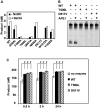
T606 and D611 are essential for ROS1 DNA glycosylase activity. (A) The generation of incision products was measured by incubating purified WT ROS1 or mutant variants (20 nM) at 30°C for 2 h with a double-stranded oligonucleotide substrate (20 nM) containing a single 5-meC:G pair. Samples were treated with or without NaOH 100 mM, and immediately transferred to 90°C for 10 min. Products were separated in a 12% denaturing polyacrylamide gel and the amounts of incised oligonucleotide were quantified by fluorescent scanning. (B) Purified WT ROS1 or mutant variants (20 nM) were incubated at 30°C for 2 h with a double-stranded oligonucleotide substrate (20 nM) containing a single 5-meC:G pair, either in the absence or the presence of human APE I (5 U), as indicated. Products were separated in a 12% denaturing polyacrylamide gel and the incised products were detected by fluorescent scanning. (C) A double-stranded oligonucleotide substrate containing an AP site opposite G (200 nM) was incubated at 30°C either in the absence of enzyme or in the presence of purified WT ROS1, T606L or D611V (100 nM). Reactions were stopped at the indicated times, products were separated in a 12% denaturing polyacrylamide gel and the amount of incised oligonucleotide was quantified by fluorescent scanning. Values are means ± SE (error bars) from two independent experiments. The asterisks indicate that the incision levels were significantly different (P < 0.05) from those observed in the absence of enzyme. The respective P-values were calculated using a Student’s unpaired t-test.
Figure 4.
Figure 4.
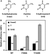
F589 and Y1028 contribute to 5-meC specificity. (A) Chemical structures of substrate DNA bases tested. (B) Substrate processing ability of WT ROS1 and the mutant variants F589A and Y1028S. Relative processing efficiencies were determined in kinetic assays as described in ‘Materials and Methods’ section. Purified proteins (20 nM), were incubated at 30°C with 51-mer double-stranded oligonucleotide substrates (20 nM) containing at position 29 of the labeled upper-strand different target DNA bases paired with G. Reaction products were separated in a 12% denaturing polyacrylamide gel and quantified by fluorescence scanning. Values are means ± SE (error bars) from two independent experiments.
Figure 5.
Figure 5.
Q607 is required for stable ROS1 binding to substrate and product DNA. Purified WT ROS1 or mutant variant Q607A (100 nM) were incubated with a mixture of 100 nM fluorescein-labeled 5-meC:G substrate and 100 nM alexa-labeled 1-nt gapped product, and the reactions were monitored for 60 min. After non-denaturing gel electrophoresis, the gel was scanned to detect fluorescein- (upper panel) or alexa-labeled (lower panel) DNA. Protein–DNA complexes were identified by their retarded mobility compared with that of free DNA, as indicated. The asterisk depicts 5′-end labeling of the upper strand. M: 5-meC.
Figure 6.
Figure 6.
N608 contributes to sequence-context specificity. (A) Purified WT ROS1 or mutant variants (20 nM) were incubated at 30°C for 4 h with 51-mer double-stranded oligonucleotide substrates (20 nM) containing at position 29 of the labeled upper-strand a 5-meC residue in different sequence contexts. Products were separated in a 12% denaturing polyacrylamide gel and the amount of incised oligonucleotide was quantified by fluorescent scanning. For ease of comparison, the incision values for each substrate are normalized to the total incision detected in all four substrates for each individual enzyme. (B) Substrate processing ability of type ROS1 and the mutant variant N608 in different sequence contexts. Relative processing efficiencies were determined in kinetic assays as described in ‘Materials and Methods’ section. Purified proteins (20 nM), were incubated at 30°C with 51-mer double-stranded oligonucleotide substrates (20 nM) containing at position 29 of the labeled upper-strand a 5-meC residue in different sequence contexts. Reaction products were separated in a 12% denaturing polyacrylamide gel and quantified by fluorescence scanning. Values are means ± SE (error bars) from two independent experiments.

Similar articles

Cited by

References

    1. Zemach A, McDaniel IE, Silva P, Zilberman D. Genome-wide evolutionary analysis of eukaryotic DNA methylation. Science. 2010;328:916–919. - PubMed
    1. Feng S, Cokus SJ, Zhang X, Chen PY, Bostick M, Goll MG, Hetzel J, Jain J, Strauss SH, Halpern ME, et al. Conservation and divergence of methylation patterning in plants and animals. Proc. Natl Acad. Sci. USA. 2010;107:8689–8694. - PMC - PubMed
    1. Roldan-Arjona T, Ariza RR. DNA demethylation. In: Grosjean H, editor. DNA and RNA modification Enzymes: Comparative Structure, Mechanism, Functions, Cellular Interactions and Evolution. Austin, TX: Landes Bioscience; 2009. pp. 149–161.
    1. Zhu JK. Active DNA demethylation mediated by DNA glycosylases. Annu. Rev. Genet. 2009;43:143–166. - PMC - PubMed
    1. Robertson KD. DNA methylation and human disease. Nat. Rev. Genet. 2005;6:597–610. - PubMed

Publication types

MeSH terms