. 2011 Jan 7;286(1):24-34.
doi: 10.1074/jbc.M110.135921. Epub 2010 Oct 29.

Bryostatin-1, a naturally occurring antineoplastic agent, acts as a Toll-like receptor 4 (TLR-4) ligand and induces unique cytokines and chemokines in dendritic cells

Affiliations

Bryostatin-1, a naturally occurring antineoplastic agent, acts as a Toll-like receptor 4 (TLR-4) ligand and induces unique cytokines and chemokines in dendritic cells

Maria Eugenia Ariza et al. J Biol Chem. .

Abstract

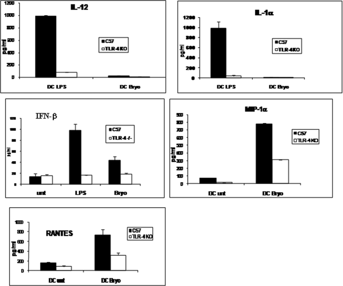
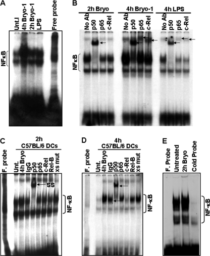
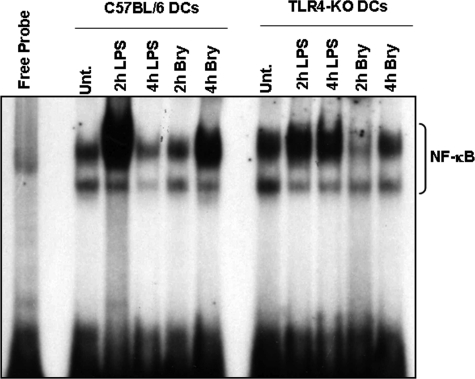
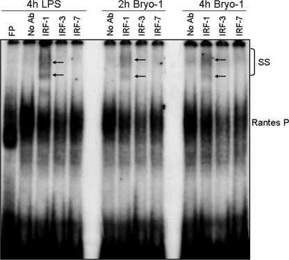
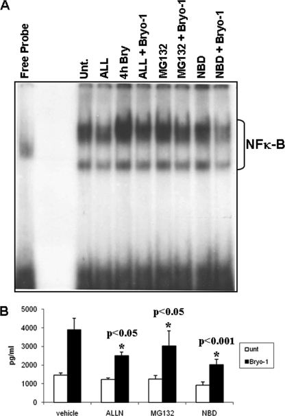
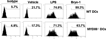
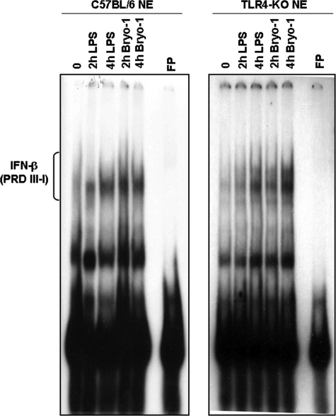
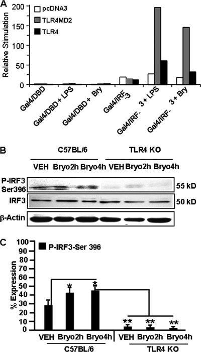
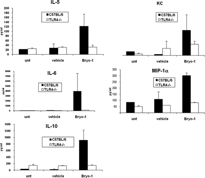
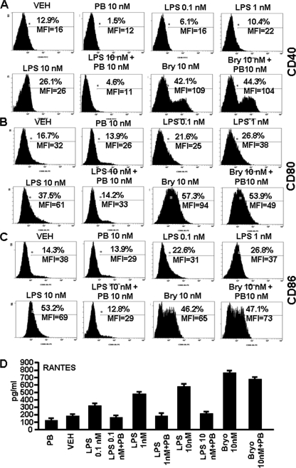
Bryostatin-1 (Bryo-1), a natural macrocyclic lactone, is clinically used as an anti-cancer agent. In this study, we demonstrate for the first time that Bryo-1 acts as a Toll-like receptor 4 (TLR4) ligand. Interestingly, activation of bone marrow-derived dendritic cells (in vitro with Bryo-1) led to a TLR4-dependent biphasic activation of nuclear factor-κB (NF-κB) and the unique induction of cytokines (IL-5, IL-6, and IL-10) and chemokines, including RANTES (regulated on activation normal T cell expressed and secreted) and macrophage inflammatory protein 1α (MIP1-α). In addition, EMSA demonstrated that Bryo-1-mediated induction of RANTES was regulated by NF-κB and the interferon regulatory factors (IRF)-1, IRF-3, and IRF-7 to the RANTES independently of myeloid differentiation primary response gene-88 (MyD88). Bryo-1 was able to induce the transcriptional activation of IRF-3 through the TLR4/MD2-dependent pathway. In vivo administration of Bryo-1 triggered a TLR-4-dependent T helper cell 2 (Th2) cytokine response and expanded a subset of myeloid dendritic cells that expressed a CD11c(high)CD8α(-) CD11b(+)CD4(+) phenotype. This study demonstrates that Bryo-1 can act as a TLR4 ligand and activate innate immunity. Moreover, the ability of Bryo-1 to trigger RANTES and MIP1-α suggests that Bryo-1 could potentially be used to prevent HIV-1 infection. Finally, induction of a Th2 response by Bryo-1 may help treat inflammatory diseases mediated by Th1 cells. Together, our studies have a major impact on the clinical use of Bryo-1 as an anti-cancer and immunopotentiating agent.


Figures

FIGURE 1.
FIGURE 1.
Bryo-1 acts as a TLR-4 ligand and triggers cytokine and chemokine production in murine BMDCs, which is significantly different from that induced by LPS. Supernatants from BMDCs from C57BL/6 wild-type or TLR4−/− mice treated with vehicle, LPS, or Bryo-1 for 24 h were examined for cytokines and chemokines as described under “Experimental Procedures.”
FIGURE 2.
FIGURE 2.
Effect of Bryo-1 on the activation of NF-κB in BMDCs. A, BMDCs from C57BL/6 mice were treated with Bryo-1 (10 ng/ml) or LPS (100 ng/ml). At the indicated time points, NF-κB DNA binding activity in nuclear extracts was determined by EMSA. Unt, untreated. B, specific formation of complexes, supershifted by anti-p50, anti-p65, and anti-c-Rel antibodies, are indicated by arrows. C and D, mutated oligonucleotide did not compete out the NF-κB complexes. F probe, free probe. E, competition assays demonstrated competing out of labeled NF-κB complexes in the presence of unlabeled wild-type NF-κB oligonucleotides.
FIGURE 3.
FIGURE 3.
Bryo-1-induced activation of NF-κB is TLR4-mediated. BMDCs from C57BL/6 or TLR4−/− mice were treated with Bryo-1 (10 ng/ml) or LPS (100 ng/ml). At the indicated time points, NF-κB DNA binding activity in nuclear extracts was determined by EMSA. Activated complexes were examined by autoradiography. Unt., untreated.
FIGURE 4.
FIGURE 4.
Bryo-1 stimulation of BMDCs induces nuclear binding of IRF-1 to the promoter of RANTES gene. Nuclear extracts were prepared 2 and 4 h after Bryo-1 or LPS (4 h) treatment, and the presence of RANTES-IRF-1 binding activity was determined by EMSA. Complex formations (supershifted (SS) band) following incubation with anti-IRF-1, anti-IRF-3, and anti-IRF-7 were visualized by autoradiography and are indicated by arrows. FP, free probe.
FIGURE 5.
FIGURE 5.
Effect of NF-κB inhibitors on Bryo-1-induced NF-κB binding activity. BMDCs were incubated for 2 h with the NF-κB inhibitors, ALL (10 μm), MG132 (1 μm), and NBD peptide (200 μm), prior to Bryo-1 stimulation for 4 h and the NF-κB binding activity in the nuclear extracts was examined by EMSA and detected by autoradiography (A). The supernatants were assayed for the presence of RANTES by a sandwich ELISA (B). Vertical bars in B represent mean ± S.E. of three independent experiments, and asterisks represent statistically significant (p < 0.05) suppression of RANTES expression in the presence of NF-κB inhibitors. Unt, untreated.
FIGURE 6.
FIGURE 6.
Production of RANTES by DCs from MyD88−/− mice. BMDCs from C57BL/6 wild-type or MyD88−/− mice treated with vehicle, LPS, or Bryo-1 for 24 h in vitro and stained with Abs against RANTES and the cells were examined by flow cytometry.
FIGURE 7.
FIGURE 7.
Induction of DNA binding activity and IFN-β production by Bryo-1 stimulation of BMDCs. Nuclear extracts were isolated from wild-type C57BL/6 or TLR4−/− mice treated with either Bryo-1 (10 ng/ml) or LPS (100 ng/ml) for the indicated times and subjected to EMSA using a 32P-labeled IFN-stimulated response element consensus sequence of the IFN-β gene promoter as probe. Activated complexes were detected by autoradiography. FP, free probe.
FIGURE 8.
FIGURE 8.
Effect of Bryo-1 on IRF-3 activation. A, pcDNA3, TLR4MD2-, and TLR4-HEK293-expressing cells were seeded in 96-well plates. After 24 h, cells were transfected with a luciferase reporter gene containing the upstream Gal4-activating sequence and Gal4/DBD (control) or Gal4/IRF-3 (50 ng) plus pCMV-β-Gal (10 ng). After 24 h, cells were stimulated with Bryo-1 (Bry) (10 ng/ml), LPS (100 ng/ml), or left untreated for 8 h, and luciferase reporter gene activity was measured. The relative stimulation value represents the ratio of firefly to β-galactosidase luciferase activities. B and C, protein from BMDCs (wild type or TLR4 KO) treated with vehicle or Bryo-1 (2 and 4 h) were fractionated in polyacrylamide gel, and expression of total IRF3 and p-IRF3-Ser-396 was analyzed by Western blotting using antibody against mouse-specific IRF3 and p-IRF3-Ser-396. Expression of p-IRF3-Ser-396 is presented as percentage of β-actin expression on the y axis, and expression of β-actin was considered to be 100%. Vertical bars represent mean ± S.E. of three independent experiments, and * represents statistically significant (p < 0.05) up-regulation of phosphorylated IRF3-Ser-396 in Bryo-1-treated groups when compared with vehicle (VEH), and ** represents statistically significant (p < 0.05) down-regulation in expression of phosphorylated IRF3-Ser-396 when Bryo-1 groups are compared between wild-type, and TLR4 KO cells are compared.
FIGURE 9.
FIGURE 9.
Bryo-1 administration in vivo triggers chemokines and Th2 cytokines in WT but not TLR4−/− mice. Serum samples from WT and TLR4−/− mice either untreated or treated with vehicle or Bryo-1 (75 μg/kg body weight) were collected 24 h post-treatment. The serum was assayed for various cytokines/chemokines by bioplex assay.
FIGURE 10.
FIGURE 10.
Effect of Bryo-1 administration in vivo on DC subpopulations in spleen. C57BL/6 mice received intraperitoneal injection of vehicle or Bryo-1, and 24 h later, the spleen cells were analyzed for the expression of CD11c, CD11b, or CD4 using flow cytometry. A and B show double staining for CD11c and CD11b, and CD11c and CD4, respectively. C depicts the total cellularity of the CD11c+CD11b+ and CD11c+CD4+ DCs in the spleens of vehicle or Bryo-1-treated C57BL/6 mice. The data represent means ± S.E. of three mice/group. The total number of cells in the Bryo-1-treated mice were significantly higher than the vehicle groups (p < 0.05).
FIGURE 11.
FIGURE 11.
DC maturation and RANTES production are Bryo-1-specific. DCs generated from bone marrow of C57BL/6 mice were cultured in the presence of vehicle (VEH), PB, Bryo-1 (Bry), Bryo-1 + PB, LPS, or LPS + PB for 24 h in vitro. The DCs were collected and analyzed by flow cytometry after staining with mouse FITC-CD40-, PE-CD80-, or PE-CD86-specific mAbs. Supernatants of various cultures were collected and analyzed for the presence of RANTES by ELISA. A–C show the expression levels of cell surface markers CD40, CD80, and CD86, respectively. The data are expressed as the mean fluorescence intensity (MFI) and represent one of three independent experiments. D represents the production of RANTES by DCs, and data are expressed as the mean levels (pg/ml) ± S.E. of triplicate cultures. The production of RANTES in the Bryo-1-treated DCs culture was significantly higher than in the vehicle group (p < 0.05). Presence of PB in the cultures containing Bryo-1 showed no significant blocking of RANTES, whereas PB caused significant inhibition of RANTES secretion in cultures with LPS (p < 0.05).

References

    1. Montoya C. J., Jie H. B., Al-Harthi L., Mulder C., Patiño P. J., Rugeles M. T., Krieg A. M., Landay A. L., Wilson S. B. (2006) J. Immunol. 177, 1028–1039 - PubMed
    1. Banchereau J., Briere F., Caux C., Davoust J., Lebecque S., Liu Y. J., Pulendran B., Palucka K. (2000) Annu. Rev. Immunol. 18, 767–811 - PubMed
    1. Zenke M., Hieronymus T. (2006) Trends Immunol. 27, 140–145 - PubMed
    1. Medzhitov R., Janeway C., Jr. (2000) Trends Microbiol. 8, 452–456 - PubMed
    1. Kaisho T., Akira S. (2001) Trends Immunol. 22, 78–83 - PubMed

Publication types

MeSH terms