

Structure-function analysis of the yeast mitochondrial Rho GTPase, Gem1p: implications for mitochondrial inheritance
- PMID: 21036903
- PMCID: PMC3012993
- DOI: 10.1074/jbc.M110.180034
Structure-function analysis of the yeast mitochondrial Rho GTPase, Gem1p: implications for mitochondrial inheritance
Abstract
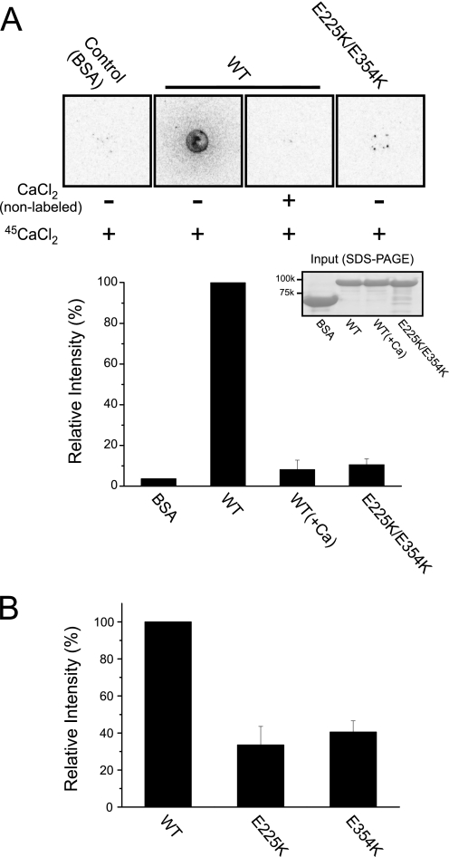
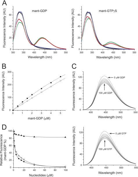
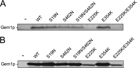
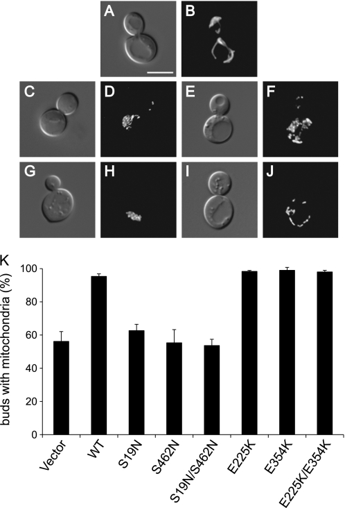
Mitochondria undergo continuous cycles of homotypic fusion and fission, which play an important role in controlling organelle morphology, copy number, and mitochondrial DNA maintenance. Because mitochondria cannot be generated de novo, the motility and distribution of these organelles are essential for their inheritance by daughter cells during division. Mitochondrial Rho (Miro) GTPases are outer mitochondrial membrane proteins with two GTPase domains and two EF-hand motifs, which act as receptors to regulate mitochondrial motility and inheritance. Here we report that although all of these domains are biochemically active, only the GTPase domains are required for the mitochondrial inheritance function of Gem1p (the yeast Miro ortholog). Mutations in either of the Gem1p GTPase domains completely abrogated mitochondrial inheritance, although the mutant proteins retained half the GTPase activity of the wild-type protein. Although mitochondrial inheritance was not dependent upon Ca(2+) binding by the two EF-hands of Gem1p, a functional N-terminal EF-hand I motif was critical for stable expression of Gem1p in vivo. Our results suggest that basic features of Miro protein function are conserved from yeast to humans, despite differences in the cellular machinery mediating mitochondrial distribution in these organisms.
Figures


Similar articles
-
Yeast Miro GTPase, Gem1p, regulates mitochondrial morphology via a novel pathway.J Cell Biol. 2004 Oct 11;167(1):87-98. doi: 10.1083/jcb.200405100. J Cell Biol. 2004. PMID: 15479738 Free PMC article.
-
A hunt for OM45 synthetic petite interactions in Saccharomyces cerevisiae reveals a role for Miro GTPase Gem1p in cristae structure maintenance.Microbiologyopen. 2021 Oct;10(5):e1238. doi: 10.1002/mbo3.1238. Microbiologyopen. 2021. PMID: 34713605 Free PMC article.
-
Multiple pathways influence mitochondrial inheritance in budding yeast.Genetics. 2008 Feb;178(2):825-37. doi: 10.1534/genetics.107.083055. Epub 2008 Feb 1. Genetics. 2008. PMID: 18245340 Free PMC article.
-
Mitochondrial manoeuvres: latest insights and hypotheses on mitochondrial partitioning during mitosis in Saccharomyces cerevisiae.Bioessays. 2010 Dec;32(12):1040-9. doi: 10.1002/bies.201000083. Epub 2010 Sep 30. Bioessays. 2010. PMID: 20886527 Review.
-
Miro (Mitochondrial Rho GTPase), a key player of mitochondrial axonal transport and mitochondrial dynamics in neurodegenerative diseases.Mitochondrion. 2021 Jan;56:118-135. doi: 10.1016/j.mito.2020.10.005. Epub 2020 Oct 28. Mitochondrion. 2021. PMID: 33127590 Review.
Cited by
-
In Candida glabrata, ERMES Component GEM1 Controls Mitochondrial Morphology, mtROS, and Drug Efflux Pump Expression, Resulting in Azole Susceptibility.J Fungi (Basel). 2023 Feb 10;9(2):240. doi: 10.3390/jof9020240. J Fungi (Basel). 2023. PMID: 36836353 Free PMC article.
-
Miro2 tethers the ER to mitochondria to promote mitochondrial fusion in tobacco leaf epidermal cells.Commun Biol. 2020 Apr 3;3(1):161. doi: 10.1038/s42003-020-0872-x. Commun Biol. 2020. PMID: 32246085 Free PMC article.
-
Insight into human Miro1/2 domain organization based on the structure of its N-terminal GTPase.J Struct Biol. 2020 Dec 1;212(3):107656. doi: 10.1016/j.jsb.2020.107656. Epub 2020 Oct 24. J Struct Biol. 2020. PMID: 33132189 Free PMC article.
-
GTP-binding protein-like domain of AGAP1 is protein binding site that allosterically regulates ArfGAP protein catalytic activity.J Biol Chem. 2012 May 18;287(21):17176-17185. doi: 10.1074/jbc.M111.334458. Epub 2012 Mar 27. J Biol Chem. 2012. PMID: 22453919 Free PMC article.
-
Make or break for mitochondria.Elife. 2013 May 14;2:e00804. doi: 10.7554/eLife.00804. Elife. 2013. PMID: 23682317 Free PMC article.
References
-
- Wang X. (2001) Genes Dev. 15, 2922–2933 - PubMed
-
- Raha S., Robinson B. H. (2000) Trends Biochem. Sci. 25, 502–508 - PubMed
-
- Rutter G. A., Rizzuto R. (2000) Trends Biochem. Sci. 25, 215–221 - PubMed
-
- Seth R. B., Sun L., Ea C. K., Chen Z. J. (2005) Cell 122, 669–682 - PubMed
-
- Moore C. B., Bergstralh D. T., Duncan J. A., Lei Y., Morrison T. E., Zimmermann A. G., Accavitti-Loper M. A., Madden V. J., Sun L., Ye Z., Lich J. D., Heise M. T., Chen Z., Ting J. P. (2008) Nature 451, 573–577 - PubMed
Publication types
MeSH terms
Substances
Grants and funding
LinkOut - more resources
Full Text Sources
Molecular Biology Databases
Miscellaneous
