. 2010 Dec;177(6):2898-911.
doi: 10.2353/ajpath.2010.100367. Epub 2010 Oct 29.

Activation of phosphatidylinositol 3-kinase/Akt and impairment of nuclear factor-kappaB: molecular mechanisms behind the arrested maturation/activation state of Leishmania infantum-infected dendritic cells

Affiliations

Activation of phosphatidylinositol 3-kinase/Akt and impairment of nuclear factor-kappaB: molecular mechanisms behind the arrested maturation/activation state of Leishmania infantum-infected dendritic cells

Bruno Miguel Neves et al. Am J Pathol. 2010 Dec.

Abstract

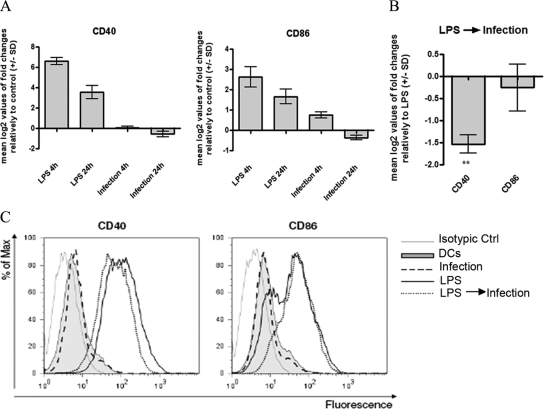
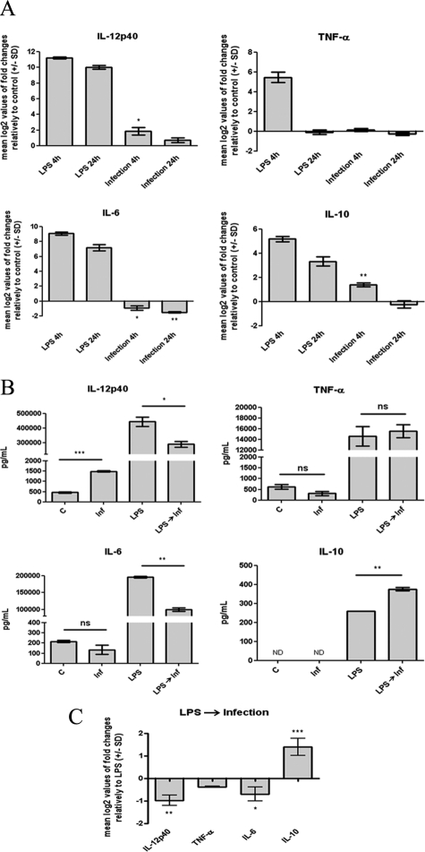
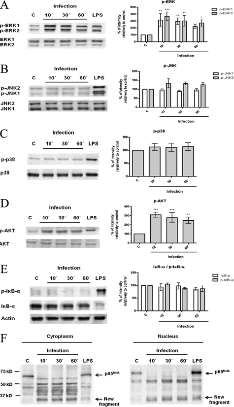
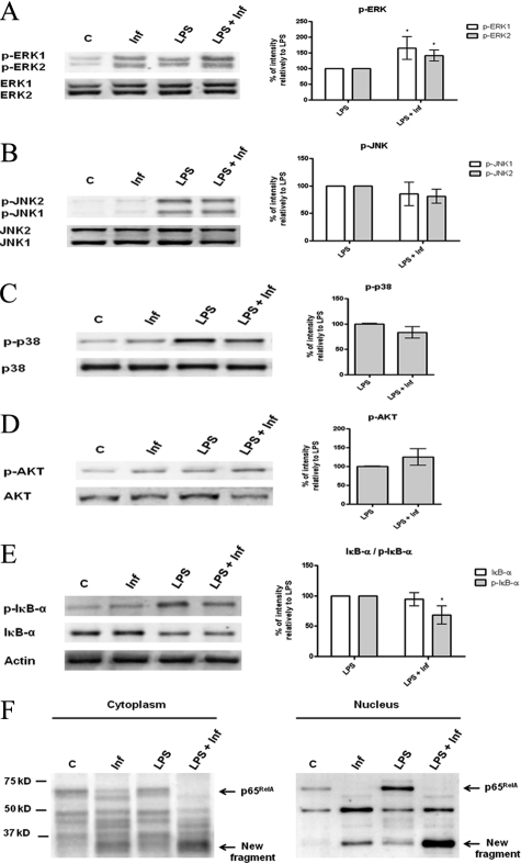
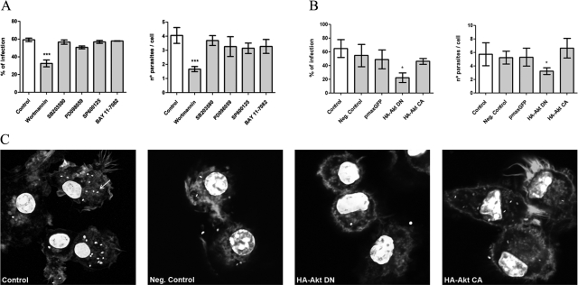
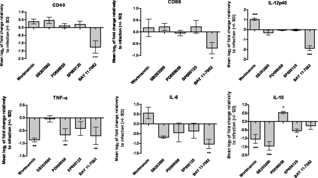
Understanding the complex interactions between Leishmania and dendritic cells (DCs) is central to the modulation of the outcome of this infection, given that an effective immune response against Leishmania is dependent on the successful activation and maturation of DCs. We report here that Leishmania infantum promastigotes successfully infect mouse bone marrow-derived DCs without triggering maturation, as shown by a failure in the up-regulation of CD40 and CD86 expression, and that parasites strongly counteract the lipopolysaccharide-triggered maturation of DCs. A small increase in interleukin (IL)-12 and IL-10 transcription and secretion and a decrease in IL-6 were observed in infected cells. This arrested DC maturation state is actively promoted by parasites because heat-killed or fixed parasites increased cytokine and costimulatory molecule expression. At a molecular level, L. infantum rapidly induced activation of phosphatidylinositol 3-kinase/Akt and extracellular signal-regulated kinase 1/2, whereas no effect was observed in the c-Jun N-terminal kinase and p38 mitogen-activated protein kinase proinflammatory pathways. Moreover, parasites actively promoted cleavage of the nuclear factor-κB p65(RelA) subunit, causing its impairment. The blockade of phosphatidylinositol 3-kinase/Akt by either treatment of bone marrow-derived DCs with wortmannin or transfection with an Akt dominant-negative mutant resulted in a strong decrease in infection rates, revealing for the first time a crucial role of this pathway on Leishmania engulfment by DCs. Overall, our data indicate that activation of Akt and impairment of nuclear factor-κB are responsible for immunogenicity subversion of L. infantum-infected DCs.


Figures

Figure 1
Figure 1
Kinetic analyses of visceral L. infantum infection. A: Cells were infected with promastigotes at a 10:1 (parasites/cell) ratio for 4 hours. BMDCs were immediately recovered or left in culture for an additional 12, 24, 48, or 72 hours. A fluorescent nuclear label, 4′,6-diamidino-2-phenylindole, was added to the cells, and the number of infected cells as well as the number of parasite per cell was counted with a fluorescence microscope at ×1000. Results are representative of three independent experiments. B: BMDCs infected with GFP-L. infantum promastigotes at a 10:1 (parasites/cell) ratio were recovered after 4 hours of incubation or left in culture for an additional 24 hours after the end of the infection. The cells were analyzed by flow cytometry, and the percentage of GFP-positive BMDCs was quantified. Each value represents the mean ± SD from three independent experiments. NS, not significant; *P < 0.05.
Figure 2
Figure 2
Effects of L. infantum infection on BMDC CD40 and CD86 costimulatory molecule expression. A: BMDCs plated at 2 × 10⁶ cells/well were infected with L. infantum promastigotes in a 10:1 (parasites/cell) ratio for 4 hours. Cells were immediately recovered or left in culture for an additional 24 hours after the end of the infection. As a control, BDMCs were stimulated with 1 μg/ml LPS for 4 and 24 hours. The mRNA levels were assessed by qPCR for CD40 and CD86. Gene expression is indicated as mean log2 values of fold changes relative to untreated cells. B: BMDCs were treated with 1 μg/ml LPS or pretreated for 1 hour with LPS and then infected with L. infantum promastigotes in a 10:1 (parasites/cell) ratio, (LPS → Infection). After 4 hours of culture, cells were harvested, and the CD40 and CD86 mRNA levels were assessed by qPCR. Gene expression is indicated as mean log2 values of fold changes relative to LPS-treated cells. C: Cells were infected, treated with 1 μg/ml LPS or pretreated during 1 hour with LPS and then infected (LPS → Infection). After 24 hours, the surface expression of CD40 and CD86 was measured on gated CD11c+ cells by flow cytometry. As negative control, cells were incubated in RPMIc (DCs) for a similar time. Each value represents the mean ± SD from three independent experiments. **P < 0.01.
Figure 3
Figure 3
Effects of L. infantum infection on BMDC cytokine production. A: Cells plated at 2 × 10⁶ cells/well were infected with L. infantum promastigotes in a 10:1 (parasites/cell) ratio or stimulated with 1 μg/ml LPS during the indicated times at 37°C with 5% CO2. The mRNA levels were assessed by real-time qPCR for IL-12p40, TNF-α, IL-6, and IL-10. Gene expression is indicated as mean log2 values of fold changes relative to untreated cells. B: The levels of IL-12p40, TNF-α, IL-6, and IL-10 were quantified by ELISA on 24-hour culture supernatants of 1 × 10⁶ BMDCs that were either infected with L. infantum promastigotes (Inf), stimulated with 1 μg/ml LPS, or pretreated for 1 hour with LPS and then infected (LPS→ Inf). C, control. C: BMDCs (2 × 10⁶/well) were stimulated with 1 μg/ml LPS or infected for 1 hour after LPS stimulation. Four hours after infection, the mRNA levels of IL-12p40, TNF-α, IL-6, and IL-10 were assessed by real-time qPCR. Gene expression is indicated as mean log2 values of fold changes relative to LPS-treated cells. Each value represents the mean ± SD from three independent experiments. *P < 0.05; **P < 0.01; ***P < 0.001; ns, not significant.
Figure 4
Figure 4
Interaction of L. infantum promastigotes with PI3K/Akt, MAPKs, and NF-κB signaling pathways. BMDCs were infected with L. infantum promastigotes in a cell/parasite ratio of 1:10, and cell lysates were prepared after 10, 30, or 60 minutes of infection. As a positive control, BMDCs were stimulated with 1 μg/ml of LPS for 30 minutes. The activation of specific intracellular signaling pathways was examined by Western blot analysis using specific antibodies to the phosphorylated (p-) forms of ERK1/2 (A), SAPK/JNK (B), p38 MAPK (C), Akt (D), and IκB-α (E). NF-κB activation was also evaluated by determination of the levels of its inhibitory protein IκB-α and by assessment of nuclear translocation of the NF-κB p65RelA subunit (F). Equal protein loading was assessed using antibodies to total ERK1/2, SAPK/JNK, p38 MAPK, and Akt or with an anti-actin antibody. The optical densities of the bands were obtained by scanning the membranes in a fluorescence scanner and then were analyzed with ImageQuant TL Software. The results are expressed as % intensity relative to control (C). Each value represents the mean ± SD from three independent experiments. *P < 0.05; **P < 0.01; ***P < 0.001.
Figure 5
Figure 5
Effects of L. infantum infection on LPS-triggered signaling pathways. BMDCs were infected with L. infantum promastigotes for 30 minutes. infantum). In some experiments, infected BMDCs were simultaneously stimulated with 1 μg/ml LPS (LPS + Inf). Nonstimulated cells (C) or LPS-stimulated cells (LPS) were used as negative and positive controls, respectively. Cell extracts were analyzed by Western blot using specific antibodies to the phosphorylated (p-) forms of ERK1/2 (A), SAPK/JNK (B), p38 MAPK (C), Akt (D), and IκB-α (E) and for total IκB-α and NF-κB p65RelA (F). Equal protein loading was evaluated with antibodies to total ERK1/2, SAPK/JNK, p38 MAPK, and Akt or with an anti-actin antibody. The optical densities of the bands were obtained by scanning the membranes in a fluorescence scanner and then were analyzed with ImageQuant TL software. The results are expressed as % intensity relative to control. Each value represents the mean ± SD from three independent experiments. *P < 0.05; **P < 0.01; ***P < 0.001.
Figure 6
Figure 6
Relevance of PI3K/Akt, MAPKs, and NF-κB signaling pathways on the engulfment of parasites by BMDCs. A: BMDCs previously treated with specific inhibitors for Akt (wortmannin), p38 MAPK (SB203580), ERK1/2 (PD098059), SAPK/JNK (SP600125), or NF-κB (Bay 11-7082) were incubated with L. infantum parasites for 4 hours. B: BMDCs were submitted to electroporation without any plasmid DNA (negative control) or transfected with pmaxGFP, a dominant-negative mutant of Akt (HA-Akt DN), or with a constitutively activated form of Akt (HA-Akt CA). After 48 hours, BMDCs were infected with L. infantum parasites. Four hours after infection, cells were stained with 4′,6-diamidino-2-phenylindole, and the numbers of infected cells as well as the numbers of parasites per cells were counted with a fluorescence microscope at ×1000. C: Images representative of fields of the different transfection experiments (original magnification: ×630) were acquired with a confocal laser scanning microscope (Zeiss LSM 510 Meta). Parasites were distributed in cytoplasm, and one of them is indicated by an arrow. Each value represents the mean ± SD from three independent experiments. *P < 0.05; **P < 0.01; ***P < 0.001.
Figure 7
Figure 7
Relevance of PI3K/Akt, MAPKs, and NF-κB signaling pathways on the expression of costimulatory molecules and cytokines by infected BMDCs. L. infantum parasites were incubated with BMDCs previously treated with specific inhibitors for Akt (wortmannin), p38 MAPK (SB203580), ERK1/2 (PD098059), SAPK/JNK (SP600125), or NF-κB (Bay 11-7082). The CD40, CD86, IL-12p40, TNF-α, IL-6, and IL-10 mRNA levels were assessed by qPCR 4 hours postinfection. Gene expression is indicated as mean log2 values of fold changes relative to L. infantum-infected cells. Each value represents the mean ± SD from three independent experiments. *P < 0.05; **P < 0.01; ***P < 0.001.

Similar articles

Cited by

References

    1. Kaye PM, Svensson M, Ato M, Maroof A, Polley R, Stager S, Zubairi S, Engwerda CR. The immunopathology of experimental visceral leishmaniasis. Immunol Rev. 2004;201:239–253. - PubMed
    1. Ghalib HW, Whittle JA, Kubin M, Hashim FA, el-Hassan AM, Grabstein KH, Trinchieri G, Reed SG. IL-12 enhances Th1-type responses in human Leishmania donovani infections. J Immunol. 1995;154:4623–4629. - PubMed
    1. Liew FY, Parkinson C, Millott S, Severn A, Carrier M. Tumour necrosis factor (TNFα) in leishmaniasis. I. TNFα mediates host protection against cutaneous leishmaniasis. Immunology. 1990;69:570–573. - PMC - PubMed
    1. Murray HW, Hariprashad J. Interleukin 12 is effective treatment for an established systemic intracellular infection: experimental visceral leishmaniasis. J Exp Med. 1995;181:387–391. - PMC - PubMed
    1. Charmoy M, Megnekou R, Allenbach C, Zweifel C, Perez C, Monnat K, Breton M, Ronet C, Launois P, Tacchini-Cottier F. Leishmania major induces distinct neutrophil phenotypes in mice that are resistant or susceptible to infection. J Leukoc Biol. 2007;82:288–299. - PubMed

Publication types

MeSH terms

Substances