. 2010 Dec;177(6):3051-60.
doi: 10.2353/ajpath.2010.100442. Epub 2010 Oct 29.

Glatiramer acetate attenuates pro-inflammatory T cell responses but does not directly protect neurons from inflammatory cell death

Affiliations

Glatiramer acetate attenuates pro-inflammatory T cell responses but does not directly protect neurons from inflammatory cell death

Alexander M Herrmann et al. Am J Pathol. 2010 Dec.

Abstract

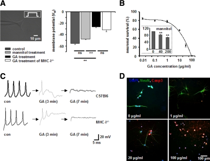
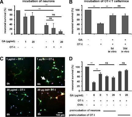
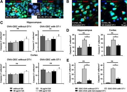
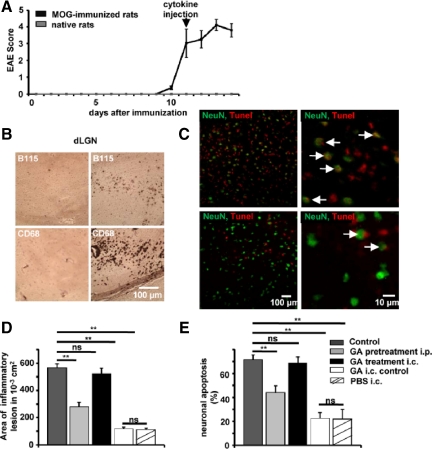
Glatiramer acetate (GA) is a synthetic, random, basic copolymer capable of modulating adaptive T cell responses. In animal models of various inflammatory and degenerative central nervous system disorders, GA-induced T cells cross the blood-brain barrier, secrete high levels of anti-inflammatory cytokines and neurotrophins, and thus both reduce neuronal damage and promote neurogenesis. Recently, it has been suggested that GA itself may permeate the (impaired) blood-brain-barrier and directly protect neurons under conditions of inflammation-mediated neurodegeneration. To test this hypothesis, we examined the direct effects of GA on neuronal functionality and T cell-mediated neuronal apoptosis in culture, acute brain slices, and focal experimental autoimmune encephalomyelitis. GA caused a depolarization of the resting membrane potential and led to an immediate impairment of action potential generation in neurons. Moreover, GA-incubated neurons underwent dose-dependent apoptosis. Apoptosis of ovalbumin peptide-loaded major histocompatibility complex class I-expressing neurons induced by ovalbumin-specific effector T cells could be reduced by pre-incubation of T cells, but not neurons with GA. Similar results could be found using acute brain slices. In focal experimental autoimmune encephalomyelitis, lesion size and neuronal apoptosis could be limited by pretreating rats with GA, whereas intracerebral GA application into the inflammatory lesion had no effect on neuronal survival. Our data suggest that GA attenuates adaptive pro-inflammatory T cell responses, but does not exert direct neuroprotective effects.


Figures

Figure 1
Figure 1
A and C: GA causes a depolarization of the resting membrane potential and an immediate impairment of action potential generation in cultured hippocampal neurons. A: Resting membrane potential of cultured WT and MHC-I−deficient hippocampal neurons in the absence or presence of GA (100 μg/ml, n = 5, respectively) and its solvent mannitol (200 μg/ml, n = 3) as determined by whole-cell patch clamp recording. C: Representative recordings of action potential firing in cultured WT (upper panels) and MHC-I-deficient hippocampal neurons without GA (con), 3 and 7 minutes after incubation with GA (100 μg/ml). Black arrows indicate the time intercept between current traces. ns = not significant, **P < 0.05. B and D: GA-incubated hippocampal neurons undergo dose-dependent apoptosis within 6 hours in culture. B: GA concentration-dependent fraction of activated caspase-3+ NeuN+ hippocampal neuronal cell bodies in culture after 6 hours of incubation. (inset) Fraction of activated caspase-3+ NeuN+ hippocampal neuronal cell bodies in culture after 6 hours of incubation with the GA solvent mannitol at different concentrations (0, 40, 200 μg/ml), ns = not significant, **P < 0.05. D: Representative immunofluorescence images of cultured hippocampal neurons exposed for 6 hours to various GA concentrations. DAPI (blue), NeuN (green), activated caspase-3 (red). Scale bar = 100 μm.
Figure 2
Figure 2
Apoptosis of OVA peptide−loaded MHC-I−expressing neurons induced by OVA-specific OT-I effector T cells is reduced by pre-incubation of OT-I splenocytes but not neurons with GA. A: Fractions of activated caspase-3+ NeuN+ cell bodies of OVA peptide−loaded hippocampal neurons in culture after 6 hours of incubation with and without in vitro activated OT-I T cells in the absence or presence of two concentrations of GA (1 and 20 μg/ml). B: Fractions of activated caspase-3+ NeuN+ cell bodies of OVA peptide−loaded hippocampal neurons in culture after 6 hours of incubation with and without activated OT-I T cells either pretreated with GA (1 or 20 μg/ml) during antigen stimulation in vitro or isolated from OT-I mice pretreated with GA (100 mg/kg/d i.p.) for 30 days. ns = not significant, **P < 0.05. C: Representative immunofluorescence images of cultured hippocampal neurons incubated for 6 hours with and without in vitro activated OT-I T cells in the absence or presence of two concentrations of GA (1 and 20 μg/ml). White arrow indicates an activated caspase-3+ NeuN+ neuron. DAPI (blue), NeuN (green), activated caspase-3 (red). Scale bar = 100 μm. D: Fractions of activated caspase-3+ NeuN+ cell bodies of non-OVA peptide−loaded hippocampal neurons in culture after 6 hours of incubation with GA (1 and 20 μg/ml) alone or with OT-I T cells activated in the absence or presence of GA (1 and 20 μg/ml). ns = not significant, **P < 0.05.
Figure 3
Figure 3
Direct oligodendroglial and collateral neuronal apoptosis in slices from ODC-OVA mice incubated with OVA-specific OT-I effector T cells is reduced by pre-incubation of OT-I splenocytes but not slices with GA. A and C: GA does not prevent neuronal cell death in acute brain slices from ODC-OVA mice on incubation for 6 hours with OT-I effector T cells. A: Representative immunofluorescence images of ODC-OVA slices incubated for 6 hours with in vitro activated OT-I T cells in the absence (left panel) and presence (right panel) of GA (50 μg/ml) in the hippocampus. (Inset) Nuclei of activated caspase-3+ neurons show inhomogeneous DAPI-staining consistent with nuclear condensation and fragmentation due to apoptotic cell death. DAPI (blue), NeuN (green), activated caspase-3 (red); scale bar represents 10 μm. C: Densities of activated caspase-3+ NeuN+ neuronal cell bodies detected in the hippocampal (upper panels) and cortical (lower panels) gray matter of acute brain slices from ODC-OVA mice incubated for 6 hours with (right panels) and without (left panels) in vitro activated OT-I T cells in the absence and presence of three different GA concentrations (10, 50, and 200 μg/ml). ns = not significant, **P < 0.05. B, D, and E: Neuronal (and oligodendroglial) apoptosis can be reduced by pre-incubation of OT-I splenocytes with GA before incubation in acute brain slices from ODC-OVA mice. B: Representative immunofluorescence images of ODC-OVA slices incubated for 6 hours with OT-I T cells in vitro activated in the absence (left panel) and presence (right panel) of GA (1 μg/ml). DAPI (blue), NeuN (green), activated caspase-3 (red). Scale bar = 10 μm. D: Densities of activated caspase-3+ NeuN+ neuronal cell bodies detected in the hippocampal (left panel) and cortical (right panel) gray matter of acute brain slices from ODC-OVA mice incubated for 6 hours without (left bar) or with OT-I T cells in vitro activated in the absence (middle bar) and presence (right bar) of GA (1 μg/ml). E: Densities of activated caspase-3+ NogoA+ oligodendrocytes detected in the hippocampal (left panel) and cortical (right panel) gray matter of acute brain slices from ODC-OVA mice incubated for 6 hours without (left bar) or with OT-I T cells in vitro activated in the absence (middle bar) and presence (right bar) of GA (1 μg/ml). ns = not significant, **P < 0.05.
Figure 4
Figure 4
In focal EAE, lesion size and neuronal apoptosis can be limited by pretreating rats, but not by i.c. application of GA into the inflammatory lesion. A: Clinical course of MOG-induced EAE in female DA rats. B: Histological analysis of the dLGN of MOG-immunized mice, at the side of i.c. cytokine injection (right panels) and the contra-lateral control side (left panels). Stainings are shown for T cells (B115, upper panels) and macrophages (CD68, lower panels). C: Representative immunofluorescence images within the dLGN of MOG-immunized rats without (upper panel) and with GA pretreatment for 30 days (lower panel). NeuN (green), TUNEL (red), and TUNEL+ neuronal cell bodies are indicated by white arrows. Scale bar = 100 or 10 μm. D and E: Quantitative analysis of lesion size and fraction of NeuN, TUNEL+ neurons within the dLGN in MOG-immunized rats 2 days after i.c. injection of pro-inflammatory cytokines (control), in MOG-immunized rats i.p. treated with GA [2 mg/d, GA pretreatment (30 days)], in MOG-immunized rats that received i.c. injection of pro-inflammatory cytokines with GA (40 μg; GA treatment i.c.) and immunized rats that received a control injection with GA (40 μg, GA i.c. control) or PBS alone. ns = not significant, **P < 0.05.

Similar articles

Cited by

References

    1. Duda PW, Schmied MC, Cook SL, Krieger JI, Hafler DA. Glatiramer acetate (Copaxone) induces degenerate. Th2-polarized immune responses in patients with multiple sclerosis. J Clin Invest. 2000;105:967–9762. - PMC - PubMed
    1. Fridkis-Hareli M, Teitelbaum D, Gurevich E, Pecht I, Brautbar C, Kwon OJ, Brenner T, Arnon R, Sela M. Direct binding of myelin basic protein and synthetic copolymer 1 to class II major histocompatibility complex molecules on living antigen-presenting cells–specificity and promiscuity. Proc Natl Acad Sci USA. 1994;91:4872–4876. - PMC - PubMed
    1. Neuhaus O, Farina C, Yassouridis A, Wiendl H, Then Bergh F, Dose T, Wekerle H, Hohlfeld R. Multiple sclerosis: comparison of copolymer-1- reactive T cell lines from treated and untreated subjects reveals cytokine shift from T helper 1 to T helper 2 cells. Proc Natl Acad Sci USA. 2000;97:7452–7457. - PMC - PubMed
    1. Weber MS, Prod'homme T, Youssef S, Dunn SE, Rundle CD, Lee L, Patarroyo JC, Stuve O, Sobel RA, Steinman L, Zamvil SS. Type II monocytes modulate T cell-mediated central nervous system autoimmune disease. Nat Med. 2007;13:935–943. - PubMed
    1. Aharoni R, Eilam R, Domev H, Labunskay G, Sela M, Arnon R. The immunomodulator glatiramer acetate augments the expression of neurotrophic factors in brains of experimental autoimmune encephalomyelitis mice. Proc Natl Acad Sci USA. 2005;102:19045–19050. - PMC - PubMed

Publication types

MeSH terms