Comparative Study
. 2010 Dec 1;185(11):6839-49.
doi: 10.4049/jimmunol.1001360. Epub 2010 Oct 29.

TIM-4, a receptor for phosphatidylserine, controls adaptive immunity by regulating the removal of antigen-specific T cells

Affiliations
Comparative Study

TIM-4, a receptor for phosphatidylserine, controls adaptive immunity by regulating the removal of antigen-specific T cells

Lee A Albacker et al. J Immunol. .

Abstract

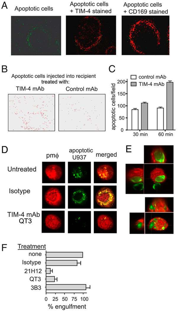
Adaptive immunity is characterized by the expansion of an Ag-specific T cell population following Ag exposure. The precise mechanisms, however, that control the expansion and subsequent contraction in the number of Ag-specific T cells are not fully understood. We show that T cell/transmembrane, Ig, and mucin (TIM)-4, a receptor for phosphatidylserine, a marker of apoptotic cells, regulates adaptive immunity in part by mediating the removal of Ag-specific T cells during the contraction phase of the response. During Ag immunization or during infection with influenza A virus, blockade of TIM-4 on APCs increased the expansion of Ag-specific T cells, resulting in an increase in secondary immune responses. Conversely, overexpression of TIM-4 on APCs in transgenic mice reduced the number of Ag-specific T cells that remained after immunization, resulting in reduced secondary T cell responses. There was no change in the total number of cell divisions that T cells completed, no change in the per cell proliferative capacity of the remaining Ag-specific T cells, and no increase in the development of Ag-specific regulatory T cells in TIM-4 transgenic mice. Thus, TIM-4-expressing cells regulate adaptive immunity by mediating the removal of phosphatidylserine-expressing apoptotic, Ag-specific T cells, thereby controlling the number of Ag-specific T cells that remain after the clearance of Ag or infection.


Conflict of interest statement

Disclosures

The authors have no financial conflicts of interest.

Figures

FIGURE 1
FIGURE 1
Blocking TIM-4 inhibits engulfment of apoptotic cells. A, Apoptotic thymocytes were labeled with the pH-sensitive dye pHrodo, incubated with untransfected or mTIM-4–transfected 3T3 cells for 90 min, and then washed. The engulfment of apoptotic cells from cell surface (weakly fluorescent at neutral pH) into acidic cell compartments (strongly fluorescent at acidic pH) was determined by flow cytometry. One experiment representative of three is shown. B, mTIM-4–transfected 3T3 cells were untreated or pretreated with TIM-4 mAb or control mAb for 15 min, incubated with CMFDA-labeled apoptotic cells for 90 min, and analyzed by FACS. Data are presented as percentage of 3T3 cells that have engulfed an apoptotic cell compared with control. C, Control 3T3 cells and mTIM-4–transfected 3T3 cells were incubated with pHrodo-labeled apoptotic thymocytes (AT) or without apoptotic thymocytes (no AT) for 30 min. TIM-4–3T3 cells were pretreated with TIM-4 mAb (21H12) or control mAb (IgG) for 15 min prior to addition of AT. Phrodo-labeled AT were pretreated with annexin V (0.1 mg/ml) for 1 h prior to addition to 3T3–TIM-4 cells.
FIGURE 2
FIGURE 2
Blockade of TIM-4 results in increased numbers of Ag-specific T cells following immunization or influenza infection. A, BALB/c mice were treated with isotype or TIM-4 mAb and immunized with 300 µg OVA in alum. Seven days later, B-depleted splenocytes were cultured with OVA, and [3H]thymidine incorporation, IL-4, and IFN-γ production were measured. One representative experiment of three is shown. B, OVA-specific DO11.10 T cells (5 × 10⁴) were transferred to recipients that were treated as in A and enumerated (mean + SEM) on days 3, 5, 7, and 9. Pooled data from two experiments shown. C, OVA-specific DO11.10 T cells from B were analyzed for expression of CD44 and CD62L on days 5 and 9. Representative FACS plots from day 9 are shown. D, Mean fluorescence intensity (MFI) of CD44 and percentage of CD62L low cells were quantified. E, OVA-specific DO11.10 T cells were transferred into recipients on day 0, and recipients were treated with TIM-4 mAb or control mAb on days 4 and 7. Spleens were harvested on day 9, and the number of DO11.10 T cells per spleen was quantified. F, BALB/c mice treated with isotype or TIM-4 mAb were infected with 1.2 × 10⁴ PFU of influenza A strain Mem/71. After 5, 10, and 14 d, lungs were harvested and stained for CD45, CD8, and H-2Kd tetramer loaded with NP147–155 (TYQRTRALV). Pooled data of three experiments shown. G, Representative FACS plots are shown.
FIGURE 3
FIGURE 3
Blockade of TIM-4 delays clearance of apoptotic cells in vivo. A, CMFDA-labeled apoptotic thymocytes (green) were i.v. injected into WT BALB/c mice, and the spleen was examined 1 h after injection. Cryosections were stained with TIM-4 mAb (21H12) or with CD169 mAb (red). Images were acquired with a fluorescence microscope (original magnification ×200). B, WT BALB/c mice were treated with TIM-4 mAb or with control mAb 4 h prior to i.v. injection of CMFDA-labeled apoptotic thymocytes, and spleens were obtained 1 h later. A reverse-color image of microscope pictures is shown. C, Accumulation of CMFDA-labeled apoptotic thymocytes into spleen was quantified by counting 30 fields of 6-µm spleen sections from TIM-4 mAb- or control mAb-treated mice. D, Cell-Tracker orange-labeled PMϕs (orange) were preincubated with TIM-4 mAb or isotype mAb for 15 min, followed by coculture with CMFDA-labeled apoptotic U937 cells (green) for 30 min before confocal microscopy imaging with a 100× objective. Images of single cells are shown. E, Images of two PMϕs are shown. Apoptotic U937 cells (green) were localized inside the PMϕs (orange) by the longitudinal (left panel) and perpendicular (upper right panel) trajectories of three-dimensional confocal sections. F, PMϕs were untreated or pretreated with TIM-4 mAbs (21H12 or QT3.14), TIM-1 mAb (3B3), or isotype control for 10 min, followed by coculture with CMFDA-labeled apoptotic thymocytes for 15 min. Cells were washed and analyzed for the presence of CMFDA-positive thymocytes in PMϕs by flow cytometry. Data are presented as percentage of engulfment compared with control.
FIGURE 4
FIGURE 4
TIM-4 mediates phagocytosis of apoptotic and activated T cells. A–C, A total of 5 × 10⁴ T cells was cultured with 5 µg/ml plate-bound anti-CD3, 2 ng/ml IL-2, and 104 3T3-Vector or 3T3–TIM-4 cells and analyzed for T cell expansion. A, T cell counts with SD are shown. B, CFSE plots at 90 h show equivalent number of T cell divisions, as analyzed by the proliferation function in FlowJo. C, Forward scatter-side scatter plots show gates for BD Calibrite beads, apoptotic cells, and live cells. D, Purified CD4+ T cells were stimulated with plate-bound anti-CD3 and soluble anti-CD28 for 9 d and then labeled with CMFDA. Untransfected or mTIM-4–transfected 3T3 cells were pretreated with TIM-4 mAb or control mAb, incubated with labeled apoptotic CD4+ T cells, and analyzed by FACS. Data are presented as percentage of engulfment, indicating the percentage of transfected cells that have phagocytosed apoptotic cells. One experiment representative of three is shown.
FIGURE 5
FIGURE 5
TIM-4 Tg mice immunized with OVA demonstrate greatly reduced secondary T cell responses. A, TIM-4 Tg or WT BALB/c mice were immunized with OVA (300 µg) in IFA s.c. Lymph nodes were removed after 9 d and cells were cultured with OVA, as indicated. B, WT or TIM-4 Tg mice were immunized three times with 10 µg OVA in alum i.p. on days 0, 14, and 21. On day 26, mice were sacrificed and splenocytes were restimulated with OVA in vitro. C, WT and TIM-4 Tg mice were immunized on days 0 and 14 with saline or 50 µg OVA in alum i.p. On days 14 and 24–27, mice received 50 µg OVA or saline intranasally. Airway resistance was measured using invasive BUXCO. One representative experiment of three is shown. Statistical analysis was performed using a two-way ANOVA with Bonferoni post-test to compare WT and Tg OVA groups, n = 5. D and E, WT and TIM-4 Tg mice were immunized with 50 µg OVA in alum i.p. (D) or in CFA i.p. (E). Spleens were removed after 9 d, and CD4+ T cells were purified and cultured with OVA and irradiated splenocytes from a WT mouse. Results are shown as the mean ± SD. ** p < 0.01.
FIGURE 6
FIGURE 6
Following secondary immunization, TIM-4 Tg mice have reduced numbers of Ag-specific T cells without alteration in function or generation of Foxp3+ Treg. A, CD4+ T cells purified from TCR Tg DO11.10 Rag−/− mice were adoptively transferred (105 cells/mouse) to WT or TIM-4 Tg mice. Mice were immunized with OVA (50 µg) in alum, and 9 d later the percentage of CD4+Foxp3+ Treg cells in the popliteal lymph node was determined by flow cytometry by first gating on TCRβ+ cell population. Two representative mice of four are shown. B, DO11.10 T cells were CFSE labeled and adoptively transferred (3 × 10⁶ cells /mouse) to WT or TIM-4 Tg mice. Recipients were immunized after 15 h with OVA (50 µg) in alum i.p. Spleens were removed after 5 d, and proliferation of OVA-specific T cells was detected as dilution of CFSE, by gating on CD4+ KJ1-26+ population. One experiment of two is shown. C, WT and TIM-4 Tg mice received 105 DO11.10 Rag−/− T cells i.v., and were immunized i.p. with OVA in alum on days 1 and 8. Spleens were harvested on day 15, and the number of DO11.10-specific cells was quantified. Pooled data of two experiments are shown. D, Cells from C were stained with the TCR clonotype-specific Ab KJ1-26, CD4, TCRβ, CD44, and CD62L. CD4 and KJ1-26 plots (top panels) show cells gated on CD4+ TCRβ+ cells. E, KJ1-26+CD4+ cells from D were subgated and plotted for CD44 and CD62L. Mean fluorescent intensity of CD62L and CD44 is shown. Quantification of mean fluorescent intensity of these markers reveals no significant differences between WT and TIM-4 Tg mice (p > 0.05, Student t test, mean + SEM shown). DO11.10 T cells were not detected in control WT mice that did not receive DO11.10 T cells. F, WT and TIM-4 Tg mice received DO11.10 cells i.v. and were immunized with OVA in alum. Seven days later, DO11.10 T cells were sorted from pooled spleens of recipient mice, and 5 × 10⁴ DO11.10 cells were cultured with 5 × 10⁴ irradiated WT splenocytes pulsed with OVA 323–339 peptide. One experiment of two is shown. *p < 0.05, Student t test, mean + SEM shown.

Similar articles

Cited by

References

    1. Kaech SM, Wherry EJ. Heterogeneity and cell-fate decisions in effector and memory CD8+ T cell differentiation during viral infection. Immunity. 2007;27:393–405. - PMC - PubMed
    1. Ravichandran KS, Lorenz U. Engulfment of apoptotic cells: signals for a good meal. Nat. Rev. Immunol. 2007;7:964–974. - PubMed
    1. Borisenko GG, Matsura T, Liu SX, Tyurin VA, Jianfei J, Serinkan FB, Kagan VE. Macrophage recognition of externalized phosphatidylserine and phagocytosis of apoptotic Jurkat cells—existence of a threshold. Arch. Biochem. Biophys. 2003;413:41–52. - PubMed
    1. McIntire JJ, Umetsu SE, Akbari O, Potter M, Kuchroo VK, Barsh GS, Freeman GJ, Umetsu DT, DeKruyff RH. Identification of Tapr (an airway hyperreactivity regulatory locus) and the linked Tim gene family. Nat. Immunol. 2001;2:1109–1116. - PubMed
    1. Freeman GJ, Casasnovas JM, Umetsu DT, DeKruyff RH. TIM genes: a family of cell surface phosphatidylserine receptors that regulate innate and adaptive immunity. Immunol. Rev. 2010;235:172–189. - PMC - PubMed

Publication types

MeSH terms