. 2010 Dec 1;185(11):6911-20.
doi: 10.4049/jimmunol.1002596. Epub 2010 Oct 29.

Chlamydia-specific CD4 T cell clones control Chlamydia muridarum replication in epithelial cells by nitric oxide-dependent and -independent mechanisms

Affiliations

Chlamydia-specific CD4 T cell clones control Chlamydia muridarum replication in epithelial cells by nitric oxide-dependent and -independent mechanisms

Krupakar Jayarapu et al. J Immunol. .

Abstract

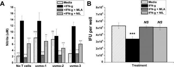
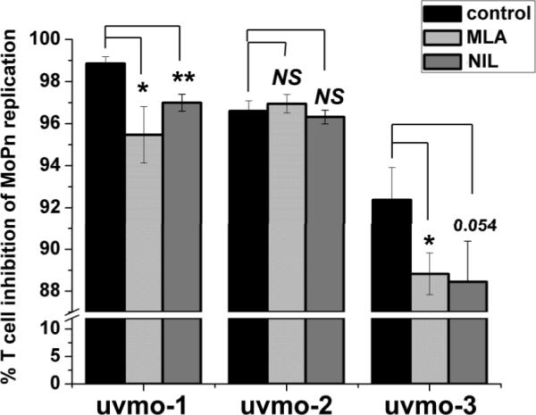
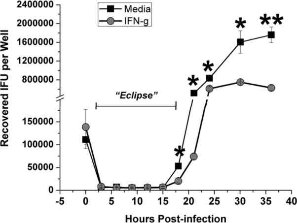
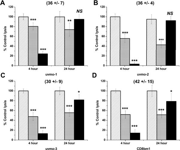
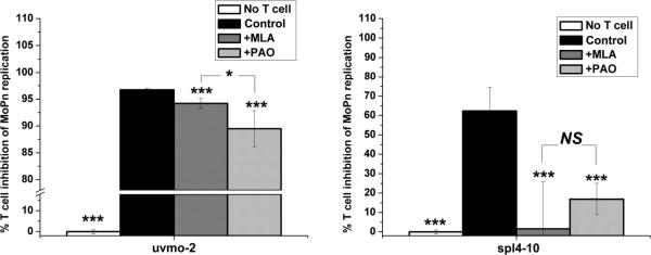
Chlamydia trachomatis serovars D-K are sexually transmitted intracellular bacterial pathogens that replicate in epithelial cells lining the human reproductive tract. It is clear from knockout mice and T cell depletion studies using Chlamydia muridarum that MHC class II and CD4 T cells are critical for clearing bacteria from the murine genital tract. It is not clear how CD4 T cells interact with infected epithelial cells to mediate bacterial clearance in vivo. Previous work using an epithelial tumor cell line showed that a Chlamydia-specific CD4 T cell clone was able to inhibit C. muridarum replication in vitro via induction of epithelial NO production. We have previously shown that Chlamydia-specific CD4 T cell clones can recognize and be activated by infected reproductive tract epithelial cells and block Chlamydia replication in them. We extend those observations by investigating the mechanism used by a panel of CD4 T cell clones to control Chlamydia replication in epithelial cells. We found that Chlamydia-specific CD4 T cell clones were cytolytic, but that cytolysis was not likely critical for controlling C. muridarum replication. For one, CD4 T cell clone-induced epithelial NO production was critical for controlling replication; however, the most potent CD4 T cell clones were dependent on T cell degranulation for replication control with only a minor additional contribution from NO production. We discuss our data as they relate to existing knockout mouse studies addressing mechanisms of T cell-mediated control of Chlamydia replication and their implications for intracellular epithelial pathogens in mouse models.


Figures

Figure 1
Figure 1
Infected epithelial cell production of nitric oxide in the absence and presence of CD4 T cell clones; C. muridarum replication in C57epi.1 epithelial cells in the absence and presence of iNOS inhibitors. A) C57epi.1 epithelial cells, untreated (hatched bars) or pretreated with IFN-γ (10 η/ml × 14 h) in the absence (black bars) or presence of iNOS inhibitors MLA (gray bars) or NIL (light gray bars), were infected with C. muridarum (3 IFU per cell). 4 h later the inocula were removed and monolayers co-cultured without and with CD4 T cell clones uvmo-1,-2,-3 for an additional 32 h. Culture supernatants were collected 36 h post infection, 32 h post co-culture, and nitric oxide quantified as nitrite; aggregate means and SEM from two independent experiments. Asterisks indicate comparisons with IFN-γ treatment (black bars) for each of the four conditions (No T cells, uvmo-1,-2,-3). Daggers indicate comparisons with the “no T cell” infected control (hatched bar). ** or†† = pvalue <0.005; ††† or = pvalue <0.0005. B) Effects of iNOS inhibitors on C. muridarum replication in infected C57epi.1 epithelial cells. C57epi.1 cells were untreated (hatched bar) or pretreated with IFN-γ (10 ηg/ml × 14 h) in absence (black bar) or presence of MLA (gray bar) or NIL (light gray bar), and then infected with C. muridarum (3 IFU per cell). Wells were harvested 36 h post infection and C. muridarum quantified on McCoy monolayers. Comparisons were made to the infected control (hatched bar). Aggregate means and SEM from two independent experiments. ††† = pvalue <0.0005; NS = not statistically significant.
Figure 2
Figure 2
IFN-γ-independent CD4 T cell clones control C. muridarum replication in epithelial cells by an iNOS-independent mechanism. C57epi.1 epithelial cells were pretreated for 14 h with IFN-γ in the absence (black bars) or presence of iNOS inhibitors MLA (light gray bars) or NIL (gray bars), then infected with 3 IFU per cell C. muridarum. Inocula were removed 4 h later and 1.5×10⁵ T cell clone cells added in the absence or presence of MLA or NIL; wells were harvested 36 h post infection and C. muridarum quantified on McCoy monolayers. Aggregate means and SEM from three independent experiments. Comparisons were made to the no inhibitor control (black bar) for each of the three T cell clones. * = pvalue < 0.05; ** = pvalue < 0.005; NS = not statistically significant. Note break in the Y-axis scale necessary to visualize effect.
Figure 3
Figure 3
C. muridarum replication time course in C57epi.1 cells. C57epi.1 cells, untreated (squares) or pretreated with 10 ηg per ml IFN-γ for 14 h (circles), were infected with ~1 IFU per cell. Wells were harvested at indicated time points and C. muridarum quantified on McCoy monolayers; mean and SD from one experiment. Note break in Y axis. * = adjusted pvalue <0.05; ** = adjusted pvalue < 0.005.
Figure 4
Figure 4
Evaluation of the cytolytic potential of Chlamydia-specific CD4 T cell clones using standard 4 h redirected lysis assays, and 24 h inhibitor stability assays in which inhibitors were pre-incubated in media for 18 h at 37°C prior to use with T cell clones in 4 h assays: A) uvmo-1, B) uvmo-2, C) uvmo-3, D) CD8bm1 (H-2Kbm1 alloantigen-specific CD8 T cell clone). 1×10⁴ Chlamydia-specific CD4 T cells or alloreactive CD8bm1 T cells were co-cultured with 1×10⁴ P815 cells in the presence of anti-CD3 antibody 145-2c11 in 4 h assays. T cells were untreated (hatched bars) or pre-incubated with CMA (light gray bars) or PAO (black bars) for 1 h before the assay. Culture supernatants were assayed for release of lactate dehydrogenase activity using a commercial non-radioactive CTL assay to determine % specific lysis. Absolute % specific lysis values for each clone absent inhibitors are shown in parentheses. Aggregate means and SEM from three independent experiments. Comparisons are made to the untreated T cell clone (hatched bars) for each of the two conditions (4 h & 24 h). * = pvalue < 0.05; ** = pvalue < 0.005; *** = pvalue < 0.0005.
Figure 5
Figure 5
Chlamydia-specific CD4 T cell clones lyse infected C57epi.1 epithelial cells in a delayed fashion: Roles of perforin and Fas/FasLigand. C57epi.1 epithelial monolayers in eight well chamber slides were mock-infected and co-cultured without and with 1×10⁵ T cells (column A), infected 18 h with 5 IFU per cell C. muridarum and co-cultured without and with T cells (column B), infected and co-cultured without and with T cells pretreated and exposed to CMA (column C), and infected co-cultured without and with T cells in the presence of monoclonal antibodies specific for Fas and FasLigand (10 μg/ml each) (column D). All wells, mock-infected or infected for 18 h, included addition of 10 μg/ml tetracycline in the co-culture media after 18 h to halt progression of the Chlamydia infection and preserve epithelial cell viability for duration of the assay. 24 h after addition of the T cells, epithelial monolayers were stained with Cytoquick and photographed at 20× magnification. Representative data from two independent experiments.
Figure 6
Figure 6
Chlamydia-specific CD4 T cell clones lyse infected C57epi.1 epithelial cells in a delayed fashion: Roles of T cell degranulation and the CD4 co-receptor. C57epi.1 epithelial monolayers in eight well chamber slides were mock-infected and co-cultured without and with 1×10⁵ T cells (column A), infected 18 h with 5 IFU per cell C. muridarum and co-cultured without and with T cells (column B), infected and co-cultured without and with T cells pretreated and exposed to PAO (column C), and infected co-cultured without and with T cells in the presence of monoclonal antibody GK1.5 specific for CD4 (10 μg/ml) (column D). All wells, mock-infected or infected for 18 h, included addition of 10 μg/ml tetracycline in the co-culture media after 18 h to halt progression of the Chlamydia infection and preserve epithelial cell viability for duration of the assay. 24 h after addition of the T cells, epithelial monolayers were stained with Cytoquick and photographed using an inverted microscope at 20× magnification. Representative data from two independent experiments.
Figure 7
Figure 7
C. muridarum replication in C57epi.1 epithelial cells in the absence and presence of inhibitors of iNOS, perforin, and T cell degranulation. Untreated C57epi.1 cells were infected with C. muridarum at 3 IFU per cell in absence (black bar) or presence of MLA (gray bar) or CMA (light gray bar) or PAO (clear hatched bar) or MLA & CMA (light gray hatched bar) or MLA & PAO (white bar). Wells were harvested 36 h post infection and C. muridarum quantified on McCoy monolayers. Comparisons were made to the infected control (black bar). Means and SD from one experiment. * = pvalue <0.05; *** = pvalue <0.0005.
Figure 8
Figure 8
Roles of iNOS, perforin, and T cell degranulation in uvmo-2- (1st column) & uvmo-3-(2nd column) mediated control of Chlamydia replication in C57epi.1 epithelial cells. C57epi.1 epithelial cells were pretreated for 14 h with IFN-γ in the absence (black bars) or presence of iNOS inhibitor MLA, and then infected with C. muridarum at 3 IFU per cell. Inocula were removed 4 h later and infected epithelial cells were co-cultured with 1.5×10⁵ T cell clone cells in the presence of MLA (gray bars), co-cultured with T cells pretreated and exposed to CMA (light gray bars), co-cultured with T cells pretreated and exposed to PAO (clear hatched bars), co-cultured with T cells pretreated and exposed to CMA in the presence of MLA (gray hatched bars), co-cultured with T cells pretreated and exposed to PAO in the presence of MLA (white bars). A sample of supernatant was collected 36 h post infection to quantify IFN-γ by ELISA (top panels); then the wells were harvested and C. muridarum quantified on McCoy monolayers (bottom panels). % T cell inhibition of MoPn replication for each treatment condition (inhibitor) was normalized to C. muridarum replication in identical parallel wells that were not co-cultured with T cells. Aggregate means and SEM from three independent experiments. Comparisons were made to the no inhibitor control (black bars) for each of the T cell clones. * = pvalue < 0.05; ** = pvalue < 0.005; *** = pvalue < 0.0005; NS = not statistically significant.
Figure 9
Figure 9
Role of iNOS and T cell degranulation in IFN-γ-dependent (spl4-10) and -independent (uvmo-2) Chlamydia-specific CD4 T cell clones. C57epi.1 epithelial cells were pretreated for 14 h with IFN-γ in the absence or presence of iNOS inhibitor MLA, and then infected with C. muridarum at 3 IFU per cell. Inocula were removed 4 h later and infected epithelial cells were co-cultured without (white bars; set as 0% inhibition) and with 1.5×10⁵ uvmo-2 T cells (first panel) or spl4-10 T cells (second panel) in the absence of inhibitors (control black bars), in the presence of MLA (gray bars), and with T cells pretreated and exposed to PAO (light gray bars). 36 h post infection the wells were harvested and C. muridarum quantified on McCoy monolayers. % T cell inhibition of MoPn replication for each treatment condition (inhibitor) was normalized to C. muridarum replication in identical parallel wells that were not co-cultured with T cells. Aggregate means and SEM from two independent experiments. Comparisons were made to the no inhibitor control (black bars) for each of the T cell clones. * = pvalue < 0.05; *** = pvalue < 0.0005; NS = not statistically significant.

Similar articles

Cited by

References

    1. CDC, editor. Sexually Transmitted Disease Surveillance, 2008. U.S. Department of Health and Human Services; Atlanta, GA: 2009.
    1. Brunham RC, Pourbohloul B, Mak S, White R, Rekart ML. The Unexpected Impact of a Chlamydia trachomatis Infection Control Program on Susceptibility to Reinfection. J Infect Dis. 2005;192:1836–1844. - PubMed
    1. Haggerty CL, Gottlieb SL, Taylor BD, Low N, Xu F, Ness RB. Risk of sequelae after Chlamydia trachomatis genital infection in women. J Infect Dis. 201(Suppl 2):S134–155. - PubMed
    1. Morrison RP, Caldwell HD. Immunity to murine chlamydial genital infection. Infect Immun. 2002;70:2741–2751. - PMC - PubMed
    1. Morrison RP, Feilzer K, Tumas DB. Gene knockout mice establish a primary protective role for major histocompatibility complex class II-restricted responses in Chlamydia trachomatis genital tract infection. Infect Immun. 1995;63:4661–4668. - PMC - PubMed

Publication types

MeSH terms