

Phosphorylation of VE-cadherin controls endothelial phenotypes via p120-catenin coupling and Rac1 activation
- PMID: 21037229
- PMCID: PMC3023264
- DOI: 10.1152/ajpheart.00650.2010
Phosphorylation of VE-cadherin controls endothelial phenotypes via p120-catenin coupling and Rac1 activation
Abstract
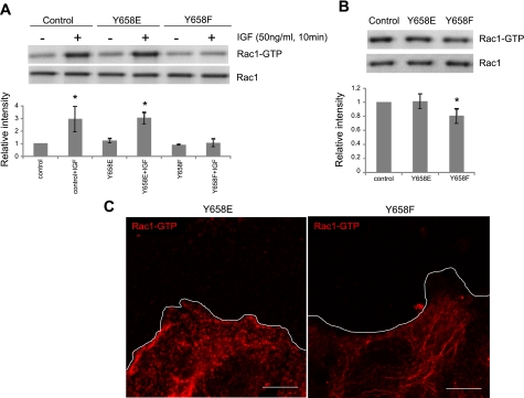
To establish the role of vascular endothelial (VE)-cadherin in the regulation of endothelial cell functions, we investigated the effect of phosphorylation of a VE-cadherin site sought to be involved in p120-catenin binding on vascular permeability and endothelial cell migration. To this end, we introduced either wild-type VE-cadherin or Y658 phosphomimetic (Y658E) or dephosphomimetic (Y658F) VE-cadherin mutant constructs into an endothelial cell line (rat fat pad endothelial cells) lacking endogenous VE-cadherin. Remarkably, neither wild-type- nor Y658E VE-cadherin was retained at cell-cell contacts because of p120-catenin preferential binding to N-cadherin, resulting in the targeting of N-cadherin to cell-cell junctions and the exclusion of VE-cadherin. However, Y658F VE-cadherin was able to bind p120-catenin and to localize at adherence junctions displacing N-cadherin. This resulted in an enhanced barrier function and a complete abrogation of Rac1 activation and lamellipodia formation, thereby inhibiting cell migration. These findings demonstrate that VE-cadherin, through the regulation of Y658 phosphorylation, competes for junctional localization with N-cadherin and controls vascular permeability and endothelial cell migration.
Figures


Similar articles
-
PKCα activation of p120-catenin serine 879 phospho-switch disassembles VE-cadherin junctions and disrupts vascular integrity.Circ Res. 2012 Aug 31;111(6):739-49. doi: 10.1161/CIRCRESAHA.112.269654. Epub 2012 Jul 12. Circ Res. 2012. PMID: 22798526 Free PMC article.
-
Unraveling the distinct distributions of VE- and N-cadherins in endothelial cells: a key role for p120-catenin.Exp Cell Res. 2010 Oct 1;316(16):2587-99. doi: 10.1016/j.yexcr.2010.06.015. Epub 2010 Jun 25. Exp Cell Res. 2010. PMID: 20599949
-
Regulation of endothelial barrier function by p120-catenin∙VE-cadherin interaction.Mol Biol Cell. 2017 Jan 1;28(1):85-97. doi: 10.1091/mbc.E16-08-0616. Epub 2016 Nov 16. Mol Biol Cell. 2017. PMID: 27852896 Free PMC article.
-
Dynamic Regulation of Vascular Permeability by Vascular Endothelial Cadherin-Mediated Endothelial Cell-Cell Junctions.J Nippon Med Sch. 2017;84(4):148-159. doi: 10.1272/jnms.84.148. J Nippon Med Sch. 2017. PMID: 28978894 Review.
-
The role of adherens junctions and VE-cadherin in the control of vascular permeability.J Cell Sci. 2008 Jul 1;121(Pt 13):2115-22. doi: 10.1242/jcs.017897. J Cell Sci. 2008. PMID: 18565824 Review.
Cited by
-
iNOS activation regulates β-catenin association with its partners in endothelial cells.PLoS One. 2012;7(12):e52964. doi: 10.1371/journal.pone.0052964. Epub 2012 Dec 28. PLoS One. 2012. PMID: 23285236 Free PMC article.
-
Agonist of growth hormone-releasing hormone reduces pneumolysin-induced pulmonary permeability edema.Proc Natl Acad Sci U S A. 2012 Feb 7;109(6):2084-9. doi: 10.1073/pnas.1121075109. Epub 2012 Jan 23. Proc Natl Acad Sci U S A. 2012. PMID: 22308467 Free PMC article.
-
Growth Differentiation Factor 6 Promotes Vascular Stability by Restraining Vascular Endothelial Growth Factor Signaling.Arterioscler Thromb Vasc Biol. 2018 Feb;38(2):353-362. doi: 10.1161/ATVBAHA.117.309571. Epub 2017 Dec 28. Arterioscler Thromb Vasc Biol. 2018. PMID: 29284606 Free PMC article.
-
A mechanistic computational model of HGF-VEGF-mediated endothelial cell proliferation and vascular permeability.iScience. 2025 Jul 24;28(8):113199. doi: 10.1016/j.isci.2025.113199. eCollection 2025 Aug 15. iScience. 2025. PMID: 40822341 Free PMC article.
-
Fibroblast growth factor signaling potentiates VE-cadherin stability at adherens junctions by regulating SHP2.PLoS One. 2012;7(5):e37600. doi: 10.1371/journal.pone.0037600. Epub 2012 May 22. PLoS One. 2012. PMID: 22629427 Free PMC article.
References
-
- Abraham S, Yeo M, Montero-Balaguer M, Paterson H, Dejana E, Marshall CJ, Mavria G. VE-Cadherin-mediated cell-cell interaction suppresses sprouting via signaling to MLC2 phosphorylation. Curr Biol 19: 668–674, 2009 - PubMed
-
- Anastasiadis PZ. p120-ctn: a nexus for contextual signaling via Rho GTPases. Biochim Biophys Acta 1773: 34–46, 2007 - PubMed
Publication types
MeSH terms
Substances
Grants and funding
LinkOut - more resources
Full Text Sources
Molecular Biology Databases
Research Materials