. 2011 Jan;300(1):H191-200.
doi: 10.1152/ajpheart.00232.2010. Epub 2010 Oct 29.

Hypotonic swelling-induced activation of PKN1 mediates cell survival in cardiac myocytes

Affiliations

Hypotonic swelling-induced activation of PKN1 mediates cell survival in cardiac myocytes

Katsuya Kajimoto et al. Am J Physiol Heart Circ Physiol. 2011 Jan.

Abstract

Hypotonic cell swelling in the myocardium is induced by pathological conditions, including ischemia-reperfusion, and affects the activities of ion transporters/channels and gene expression. However, the signaling mechanism activated by hypotonic stress (HS) is not fully understood in cardiac myocytes. A specialized protein kinase cascade, consisting of Pkc1 and MAPKs, is activated by HS in yeast. Here, we demonstrate that protein kinase N1 (PKN1), a serine/threonine protein kinase and a homolog of Pkc1, is activated by HS (67% osmolarity) within 5 min and reaches peak activity at 60 min in cardiac myocytes. Activation of PKN1 by HS was accompanied by Thr(774) phosphorylation and concomitant activation of PDK1, a potential upstream regulator of PKN1. HS also activated RhoA, thereby increasing interactions between PKN1 and RhoA. PP1 (10(-5) M), a selective Src family tyrosine kinase inhibitor, significantly suppressed HS-induced activation of RhoA and PKN1. Constitutively active PKN1 significantly increased the transcriptional activity of Elk1-GAL4, an effect that was inhibited by dominant negative MEK. Overexpression of PKN1 significantly increased ERK phosphorylation, whereas downregulation of PKN1 inhibited HS-induced ERK phosphorylation. Downregulation of PKN1 and inhibition of ERK by U-0126 both significantly inhibited the survival of cardiac myocytes in the presence of HS. These results suggest that a signaling cascade, consisting of Src, RhoA, PKN1, and ERK, is activated by HS, thereby promoting cardiac myocyte survival.


Figures
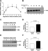
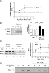
Fig. 1.
Fig. 1.
Time course of hypotonic stress-induced PKN1 activation and phosphorylation. Cardiac myocytes were subjected to hypotonic stress for the indicated periods. A: PKN1 was immunoprecipitated, and the enzyme activity was determined using the PKC-δ peptide as the substrate. The level of PKN1 kinase activity in the control state was designated as 1. Results are expressed as means ± SE obtained from 4–6 independent experiments. *P < 0.05 vs. control. B: electrophoretic mobility shift of PKN1. PKN1 was immunoprecipitated and resolved on 6% gels. In lane 6, anti-PKN1 antibody [Ab(−)] was not included in the immunoprecipitation. Molecular weight (MW) standards are indicated on the left (in kDa). Results are representative of 6 independent experiments. C: cell lysates were subjected to immunoblot analyses with anti-Thr774 phosphorylated (p-)PKN1 and anti-total PKN1 antibodies. D: Thr774 p-PKN1/total PKN1 was determined after densitometric analyses. The ratio at 60 min (hypotonic) was expressed relative to that at time 0 (control). E: cell lysates were subjected to immunoblot analyses with anti-Ser221 p-phoshoinositide-dependent protein kinase-1 (PDK1) and anti-total PDK1 antibodies. F: Ser221 p-PDK1/total PDK1 was determined after densitometric analyses. The ratio at 60 min was expressed relative to that at time 0.
Fig. 2.
Fig. 2.
Hypotonic stress-induced translocation of RhoA from the soluble fraction to the particulate fraction in cardiac myocytes. A: cardiac myocytes were stimulated with hypotonic media (67% osmolarity) for the indicated times and fractionated into soluble (lanes 1–4) and particulate (lanes 5–8) fractions. Equal amounts of protein were loaded in each lane. Immunoblot analyses were performed using specific anti-RhoA antibody. The result shown is representative of 4 independent experiments. B: the intensity of each band in the particulate fractions was determined by densitometric analyses and expressed as the value relative to that at time 0. The results are expressed as means ± SE obtained from 4 independent experiments. *P < 0.05 vs. control. C: in vitro interaction of the recombinant NH2-terminal Rho-binding domain of PKN1 [glutathione-S-transferease (GST)-PKN1 (3–135)] and RhoA. Cardiac myocytes were stimulated with hypotonic media (67% osmolarity) for the indicated times. Equal amounts of myocyte lysate were loaded onto the GST-PKN1 (3–135) column in each lane, and immunoblot analyses were performed using specific anti-RhoA antibody. Only the GTP-bound form of RhoA in the lysates bound to the GST-PKN1 (3–135) column. Bound RhoA was detected by elution of the complex with glutathione and blotting for RhoA. The results shown are representative of 5 independent experiments.
Fig. 3.
Fig. 3.
Effects of PP1, a selective Src family tyrosine kinase inhibitor, on hypotonic cell swelling-induced RhoA-PKN1 activation. Myocytes were pretreated with PP1 (10 μM) for 60 min, and hypotonic stimulation (67% osmolarity) was then applied for the indicated times. A: PKN1 kinase activity was determined by a peptide kinase assay using the PKC-δ peptide as the substrate, which was performed after immunoprecipitating PKN1 with an anti-PKN1 antibody. The level of PKN1 kinase activity in the control state was designated as 1. Results are expressed as means ± SE obtained from 3 independent experiments. PKN1 kinase activities of immunoprecipitates without pretreatment with PP1 (■) and with pretreatment with PP1 (○) are shown. *P < 0.05 vs. cells without hypotonic stress. B, left: cardiac myocytes were transduced with adenovirus harboring LacZ or dominant negative Src (DN-Src) for 48 h, and hypotonic stimulation (67% osmolarity) was then applied for 60 min. Cell lysates were subjected to immunoblot analyses with anti-Thr774 p-PKN1, anti-total PKN1, and anti-tubulin antibodies. Right, Thr774 p-PKN1/total PKN1 was determined after densitometric analyses. The value in LacZ-treated samples without hypotonic stress was expressed as 1. The results were obtained from 4 experiments. C, left: cardiac myocytes were stimulated with hypotonic media (67% osmolarity) for the indicated times and fractionated into soluble (lanes 1–3) and particulate (lanes 4–6) fractions. Equal amounts of protein were loaded in each lane. Immunoblot analyses were performed using specific anti-RhoA antibody. The results shown are representative of 4 independent experiments. Right, the intensity of each band in the particulate fraction was determined by densitometric analyses and expressed as the value relative to that at time 0. The results are expressed as means ± SE obtained from 4 independent experiments. *P < 0.05 vs. cells without hypotonic stress. D: in vitro interactions of the recombinant NH2-terminal Rho-binding domain of PKN1 [GST-PKN1 (3–135)] with RhoA were evaluated by an in vitro binding assay. Equal amounts of protein were loaded in each lane, and immunoblot analyses were performed using specific anti-RhoA antibody. Bound RhoA was detected by elution of the complex with glutathione and blotting for RhoA. In lanes 4–6, the experiments were conducted in the presence of PP1 (10 μM). The results shown are representative of 3 independent experiments.
Fig. 4.
Fig. 4.
Role of Src in mediating hypotonic stress-induced activation of PDK1. A: cardiac myocytes were treated with either PP1 (10 μM), wortmannin (Wort; 10 nM), or PP1 plus wortmannin, and hypotonic stress was then applied. Cell lysates were subjected to immunoblot analyses with anti-Ser221 p-PDK1, anti-total PDK1, anti-Thr774 p-PKN1, anti-total PKN1, and anti-tubulin antibodies. B: Ser221 p-PDK1/total PDK1 was evaluated with densitometric analyses. C: Thr774 p-PKN1/total PKN1 was determined with densitometric analyses. The results were obtained from 4 experiments. In B and C, the open bars indicate control myocytes without hypotonic or inhibitors. *P < 0.05 vs. control; #P < 0.05 vs. hypotonic stress.
Fig. 5.
Fig. 5.
Role of PKN1 in mediating the phosphorylation of Elk-1 in cardiac myocytes. A: myocytes were transfected with the Elk-1 luciferase reporter gene and pcDNA3 or constitutively active PKN1 (CA-PKN1) together with either pcDNA3 or a dominant-negative mutant of MEK1 (DN-MEK1). B: myocytes were transfected with the Elk-1 luciferase reporter gene and either pcDNA3 or kinase inactive PKN1 (KI-PKN1). Some myocytes were subjected to hypotonic stress for 3 h. In A and B, differences in the transfection efficiency were corrected by the activity of cotransfected SV40-βgal. The value of control samples transfected with pcDNA3 without stress was designated as 1. Each bar represents mean ± SE from 3 independent experiments performed in triplicate. C: cultured cardiac myocytes were transduced with adenovirus harboring PKN1 or LacZ. Forty-eight hours after transduction, cell lysates were prepared, and immunoblot analyses were then conducted with anti-p-ERK or total ERK antibody. The quantitative analyses of the immunoblot data are shown. The level of p-ERK2/ERK2 in control myocytes was expressed as 1. D: cultured cardiac myocytes were transduced with either adenovirus harboring short hairpin (sh)RNA scramble (sh-con) or shRNA-PKN1 (sh-PKN1), and, 72 h after transduction, cells were subjected to hypotonic stress for 30 min. Cell lysates were prepared, and immunoblot analyses were then conducted with anti-p-ERK or total ERK antibody. The quantitative analyses of the immunoblot data are shown. The level of p-ERK2/ERK2 in control myocytes was expressed as 1. Although we show the quantitation of ERK2 in the bar graphs, a similar trend was observed for ERK1 when the analysis was conducted with longer exposures.
Fig. 6.
Fig. 6.
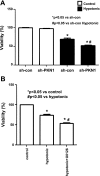
Role of PKN1 in mediating the survival of cardiac myocytes in response to hypotonic stress. A: cultured cardiac myocytes were transduced with either sh-con or sh-PKN1, and, 72 h after transduction, cells were subjected to hypotonic stress for 24 h. In A and B, the viability of cardiac myocytes was evaluated with Cell Titer Blue assays (Promega), as described in materials and methods. The viability of control myocytes without hypotonic stress was set as 100%. B: cultured cardiac myocytes were treated with U-0126 (10 μM) for 1 h and then subjected to hypotonic stress for 24 h.

Similar articles

Cited by

References

    1. Aikawa R, Komuro I, Yamazaki T, Zou Y, Kudoh S, Zhu W, Kadowaki T, Yazaki Y. Rho family small G proteins play critical roles in mechanical stress- induced hypertrophic responses in cardiac myocytes. Circ Res 84: 458–466, 1999 - PubMed
    1. Amano M, Mukai H, Ono Y, Chihara K, Matsui T, Hamajima Y, Okawa K, Iwamatsu A, Kaibuchi K. Identification of a putative target for Rho as the serine-threonine kinase protein kinase N. Science 271: 648–650, 1996 - PubMed
    1. Aoki H, Izumo S, Sadoshima J. Angiotensin II activates RhoA in cardiac myocytes:a critical role of RhoA in angiotensin II-induced premyofibril formation. Circ Res 81: 666–676, 1998 - PubMed
    1. Armstrong SC, Shivell LC, Ganote CE. Sarcolemmal blebs and osmotic fragility as correlates of irreversible ischemic injury in preconditioned isolated rabbit cardiomyocytes. J Mol Cell Cardiol 33: 149–160, 2001 - PubMed
    1. Atienza JM, Suh M, Xenarios I, Landgraf R, Colicelli J. Human ERK1 induces filamentous growth and cell wall remodeling pathways in Saccharomyces cerevisiae. J Biol Chem 275: 20638–20646, 2000 - PubMed

Publication types

MeSH terms

LinkOut - more resources