. 2010 Dec;11(12):1102-9.
doi: 10.1038/ni.1956. Epub 2010 Oct 31.

CD1a-autoreactive T cells are a normal component of the human αβ T cell repertoire

Affiliations

CD1a-autoreactive T cells are a normal component of the human αβ T cell repertoire

Annemieke de Jong et al. Nat Immunol. 2010 Dec.

Abstract

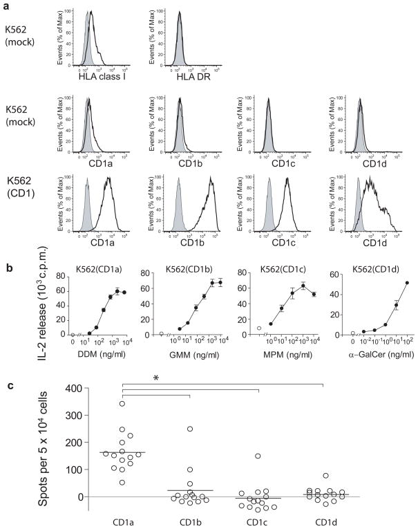
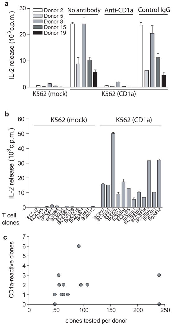
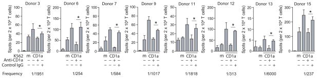
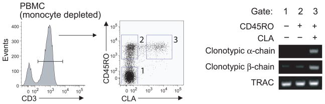
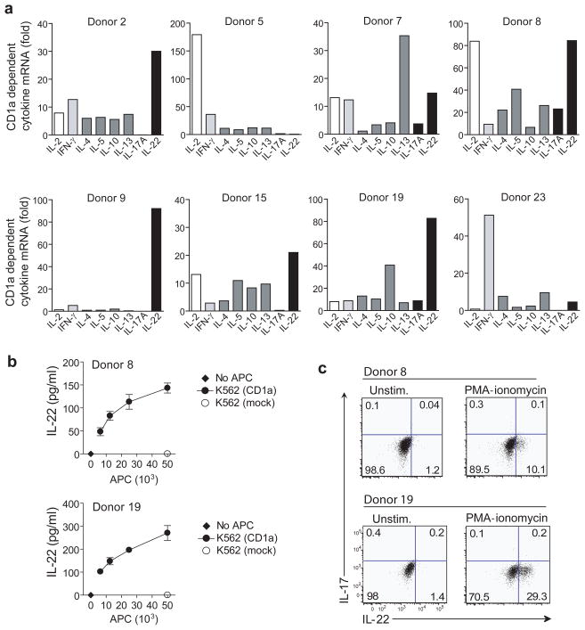
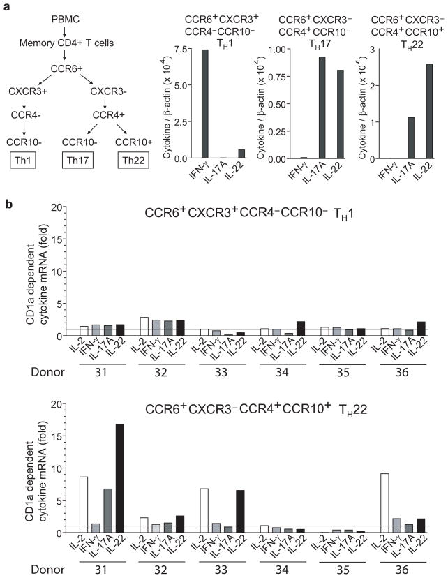
CD1 activates T cells, but the function and size of the possible human T cell repertoires that recognize each of the CD1 antigen-presenting molecules remain unknown. Using an experimental system that bypasses major histocompatibility complex (MHC) restriction and the requirement for defined antigens, we show that polyclonal T cells responded at higher rates to cells expressing CD1a than to those expressing CD1b, CD1c or CD1d. Unlike the repertoire of invariant natural killer T (NKT) cells, the CD1a-autoreactive repertoire contained diverse T cell antigen receptors (TCRs). Functionally, many CD1a-autoreactive T cells homed to skin, where they produced interleukin 22 (IL-22) in response to CD1a on Langerhans cells. The strong and frequent responses among genetically diverse donors define CD1a-autoreactive cells as a normal part of the human T cell repertoire and CD1a as a target of the T(H)22 subset of helper T cells.


Conflict of interest statement

Competing financial interests

The authors declare no competing financial interests.

Figures

Figure 1
Figure 1. A population study of CD1 autoreactive cells in blood of human donors
(a) Surface expression of MHC and CD1 in K562 cells stably transfected with vector only (mock) or with vector containing the indicated human CD1 gene. Open histograms: MHC or CD1 staining, filled histograms: Isotype control. Results are representative of three or more experiments. (b) HT2 bioassay of IL-2 release in the supernatant of K562 cells incubated for 24 h with T cell lines recognizing CD1a and dideoxymycobactin (DDM), CD1b and glucose monomycolate (GMM), CD1c and mannosyl phosphomycoketide (MPM) or CD1d and α galactosyl ceramide (mean ± S.D.). Results are representative of three or more experiments. (c) Elispot of IFNγ release in polyclonal cell cultures from 14 donors stimulated in vitro with autologous DCs and analyzed for CD1 reactivity using K562-CD1 as APCs. IFNγ release is depicted as mean number of spots after subtraction of background spots formed in response to K562 transfected with vector alone. * (P<0.01) Dunnett’s Multiple Comparison Test after one-way ANOVA.
Figure 2
Figure 2. Autoreactive T cells in the blood recognize the CD1a protein
(a) Polyclonal T cell cultures were incubated with K562 cells transfected with vector only (K562 mock), K562-CD1a cells or K562-CD1a cells pretreated with anti-CD1a blocking antibody or isotype control. After 24 hrs supernatant was analyzed for IL-2 release using HT2 bioassay (mean ± S.D.) Results are representative of three or more experiments for each T cell line. (b) T cell clones were incubated with K562 cells transfected with vector only (K562 mock) or K562-CD1a for 24 hrs and IL-2 release was measured using HT2 bioassay. Each clone was tested in two or more experiments. (c) 1291 T cell clones were generated from 14 donors by ex vivo limiting dilution and tested for CD1a response as in (b) or by incubating with K562-CD1a cells pretreated with anti-CD1a blocking antibody or isotype control and enumerating responding cells by IFN-γ Elispot assay.
Figure 3
Figure 3. Quantitative detection of CD1a-autoreactive memory T cells
CD45RO+ T cells from eight blood bank donors were incubated with K562 cells transfected with CD1a or vector only (mock, m). K562-CD1a cells were pre incubated with anti-CD1a or control IgG for 1 h before adding T cells and IFNγ-secreting cells were enumerated by Elispot assay (mean ± S.D.). * (P<0.05, Student’s t-test, two-tailed)
Figure 4
Figure 4. CD1a-autoreactive T cells in the blood express skin homing markers
RNA was extracted from CD45RO−CLA−, CD45RO+CLA−, CD45RO+CLA+ T cells and analyzed by RT PCR with primers validated to be specific for the CDR3 regions of TCR α and β chains. TCRα constant region primers (TRAC) were used as a control for input cDNA.
Figure 5
Figure 5. CD1a-dependent IL-22 production
(a) K562-CD1a cells were preincubated with anti-CD1a or control IgG and then co-cultured with polyclonal CD1a-autoreactive T cell cultures for 6 hrs at a K562 to T cell ratio of 1 : 10. CD1a dependent cytokine gene upregulation was measured by real-time PCR. Samples were normalized to β-actin. (b) T cell lines from two donors that showed upregulation of IL-22 transcripts were incubated with K562 vector only (mock) or increasing numbers of K562-CD1a. After 24 h the supernatant was analyzed for IL-22 protein by ELISA (mean ± S.D.). Results are representative of two independent experiments for each T cell line. (c) T cell lines from the same donors were analyzed for intracellular IL-17 and IL-22 staining in response to stimulation with PMA-ionomycin.
Figure 6
Figure 6. TH22 subset contains CD1a-autoreactive T cells
(a) Memory CD4+ T cells from six blood donors were sorted into CCR6+CXCR3+CCR4−CCR10−, CCR6+CXCR3−CCR4+CCR10−, and CCR6+CXCR3−CCR4+CCR10+ fractions and stimulated for 6 h with OKT3 to assess the cytokine profiles by real time PCR. (b) After a single in vitro expansion with DCs, sorted T cell fractions were co-cultured for 6 h with K562-CD1a preincubated with anti-CD1a or control IgG at a K562 to T cell ratio of 1 : 10. CD1a dependent cytokine gene upregulation was measured by real-time PCR. Samples were normalized to β-actin.
Figure 7
Figure 7. IL-22 producing CD1a-autoreactive T cells in human skin
(a) Lymphocytes from three normal human dermis samples, expanded in vitro with DC, were incubated with CD1 transfected K562 cells for 24 h after which the IL-22 release was measured by ELISA (mean ± S.D.). (b) Freshly isolated Langerhans cells, monocyte-derived DCs and K562-CD1a were incubated with a CD1a-autoreactive T cell line for 24 hrs after which supernatant was analyzed for IL-2 by HT2 bioassay (mean ± S.D.). The results are representative of separate experiments performed for three different T cell lines. The mean fluorescence intensity of CD1a on the antigen presenting cells was determined by flow cytometry. Open histograms: CD1a staining, filled histograms: Isotype control. (c) Freshly isolated Langerhans cells were pre-incubated with control IgG or anti-CD1a and the incubated with a CD1a-reactive T cell line for 18 hrs after which RNA was extracted and after reverse transcription the CD1a dependent upregulation of cytokine mRNA was measured by real time PCR. Results are representative of two experiments with natural CD1a expressing APC.

Comment in

References

    1. Fowlkes BJ, et al. A novel population of T-cell receptor alpha beta-bearing thymocytes which predominantly expresses a single V beta gene family. Nature. 1987;329:251–254. - PubMed
    1. Lantz O, Bendelac A. An invariant T cell receptor alpha chain is used by a unique subset of major histocompatibility complex class I-specific CD4+ and CD4–8− T cells in mice and humans. J Exp Med. 1994;180:1097–1106. - PMC - PubMed
    1. Porcelli S, Yockey CE, Brenner MB, Balk SP. Analysis of T cell antigen receptor (TCR) expression by human peripheral blood CD4–8− alpha/beta T cells demonstrates preferential use of several V beta genes and an invariant TCR alpha chain. J Exp Med. 1993;178:1–16. - PMC - PubMed
    1. Benlagha K, Weiss A, Beavis A, Teyton L, Bendelac A. In vivo identification of glycolipid antigen-specific T cells using fluorescent CD1d tetramers. J Exp Med. 2000;191:1895–1903. - PMC - PubMed
    1. Gumperz JE, Miyake S, Yamamura T, Brenner MB. Functionally distinct subsets of CD1d-restricted natural killer T cells revealed by CD1d tetramer staining. J Exp Med. 2002;195:625–636. - PMC - PubMed

Publication types

MeSH terms