. 2010 Dec;7(12):1003-8.
doi: 10.1038/nmeth.1526. Epub 2010 Oct 31.

Maltose-neopentyl glycol (MNG) amphiphiles for solubilization, stabilization and crystallization of membrane proteins

Affiliations

Maltose-neopentyl glycol (MNG) amphiphiles for solubilization, stabilization and crystallization of membrane proteins

Pil Seok Chae et al. Nat Methods. 2010 Dec.

Abstract

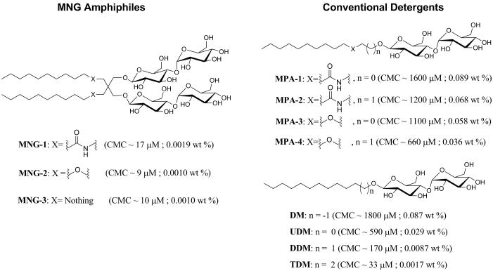
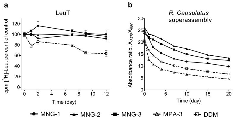
The understanding of integral membrane protein (IMP) structure and function is hampered by the difficulty of handling these proteins. Aqueous solubilization, necessary for many types of biophysical analysis, generally requires a detergent to shield the large lipophilic surfaces of native IMPs. Many proteins remain difficult to study owing to a lack of suitable detergents. We introduce a class of amphiphiles, each built around a central quaternary carbon atom derived from neopentyl glycol, with hydrophilic groups derived from maltose. Representatives of this maltose-neopentyl glycol (MNG) amphiphile family show favorable behavior relative to conventional detergents, as manifested in multiple membrane protein systems, leading to enhanced structural stability and successful crystallization. MNG amphiphiles are promising tools for membrane protein science because of the ease with which they may be prepared and the facility with which their structures may be varied.


Figures

Figure 1
Figure 1
Chemical structures of MNG amphiphiles (MNG-1, MNG-2, and MNG-3) and their linear counterparts (MPA-1, MPA-2, MPA-3, MPA-4, DM, UDM, DDM, and TDM). The critical micelle concentration (CMC) value for each agent, measured via hydrophobic dye solubilization, is indicated in parentheses.
Figure 2
Figure 2
GPCR stability in MNG amphiphiles or conventional detergents. (a) Tm values of the β2AR-T4L plotted in terms of wt % of the MNG amphiphiles (MNG-1, MNG-2, and MNG-3) or conventional detergents (MPA-1, MPA-3, DM, DDM, and TDM). β2AR-T4L with bound carazolol (an inverse agonist) was incubated with various agents at the various concentrations at indicated temperatures for 5 min prior to fluorescence emission measurements. Normalized results are expressed as mean ± s.e.m. (n = 3, 4, or 5). (b) Specific activities (pmol/mg) of M3AchR in DDM and MNG-3. The activity of the protein was evaluated after being washed and eluted with buffer including DDM or MNG-3, but without CHS, via a binding assay involving the antagonist [3H] N-methyl scopolamine, in the absence (t = 0 hr, − CHS; the first bar) or presence of CHS (t = 0 hr, + CHS; the second bar). The DDM- and MNG-3-purified M3AchR samples were stored at 4 °C for 15 hours, and then activities were measured again in the presence of CHS (t = 15 hr, + CHS; the third bar). Results are expressed as mean ± s.d. (n = 3).
Figure 3
Figure 3
SDS-12% PAGE analysis and Western blot detection of MelB. MelB samples were subjected to SDS-PAGE analysis, and MelB was detected by Western blotting using anti-His tag antibody. 10 μg membrane proteins were applied for the untreated membrane (memb.) or detergent extracts prior to ultracentrifugation (−), and equal volume of solutions were loaded for samples that the ultracentrifugation were applied (+).
Figure 4
Figure 4
Stability of SQR solubilized with MNG amphiphiles or conventional detergents. (a) CPM assays for SQR solubilized with MNG amphiphiles (MNG-1, MNG-2, and MNG-3) or conventional detergents (MPA-4, DDM, DM and SDS) at 10 × CMC. The unfolding of the each protein was monitored at 40 °C for 130 min using a microplate spectrofluorometer. Gel filtration analysis of SQR in (b) DDM or (c) MNG-3 at 10 × CMC. SQR in DDM or MNG-3 was incubated for 120 min at 40 °C. (d) Time course of SQR activity in MNG-3 or DDM. Each agent was used at 10 × CMC (0.01 wt % for MNG-3, 0.087 wt % for DDM) and 50 × CMC (0.05 wt % for MNG-3, 0.44 wt % for DDM). Note that 50 × CMC MNG-3 is comparable to DDM at 10 × CMC in terms of wt %. The catalytic rate constant (kcat) is plotted as a function of incubation time. Data at t = 0 correspond to the activity of SQR following thermal activation performed at 30 °C for 20 min. Protein solubilized with each agent was incubated at 40 °C for a further 120 min, and activity of the protein was measured at the designated times. The kcat values at each time point were calculated by analyzing reaction data according to Michaelis-Menten kinetics.
Figure 5
Figure 5
Long-term stability of LeuT and R. capsulatus superassembly in MNG amphiphiles or conventional detergents. (a) Time course of activity ([3H]-Leu binding) assay for LeuT solubilized with MNG amphiphiles (MNG-1, MNG-2, and MNG-3) and DDM at 0.026 wt % above its critical micelle concentration (CMC) (total concentrations: 0.035 wt % DDM, 0.028 wt % MNG-1, 0.027 wt % MNG-2 and 0.027 wt % MNG-3). LeuT activity was monitored at indicated time points, using a scintillation proximity assay (SPA), for protein stored at the room temperature. Results are expressed as % activity relative to the appropriate day 0 measurement. Normalized results are expressed as mean ± s.e.m. (n = 2). (b) Time course of stability of R. capsulatus superassembly purified with MNG amphiphiles (MNG-1, MNG-2, and MNG-3) and conventional detergents (MPA-3 and DDM) at 1 × CMC. The absorption ratios (A875/A680) of the detergent or amphiphile samples were followed as a function of time.
Figure 6
Figure 6
Image and x-ray diffraction pattern from crystals of cytochrome b6f /MNG-3 complexes. X-ray diffraction by cytochrome b6f crystal obtained in the presense of MNG-3. The left panel represents a portion of the pattern (0.5 degree oscillation range). Resolution limits are marked with arrows (the white cross is due to the tiling of the detector). Top right: enlargement of the red square with two strong spots near the resolution limit. A section through the two strong spots is shown in the lower right corner.

References

    1. Liu J, Rost B. Comparing function and structure between entire proteomes. Protein Sci. 2001;10:1970–1979. - PMC - PubMed
    1. Lacapere JJ, Pebay-Peyroula E, Neumann JM, Etchebest C. Determining membrane protein structures: still a challenge! Trends Biochem. Sci. 2007;32:259–270. - PubMed
    1. Privé GG. Detergents for the stabilization and crystallization of membrane proteins. Methods. 2007;41:388–397. - PubMed
    1. Schafmeister CE, Meircke LJW, Stroud RM. Structure at 2.5 Å of a designed peptide that maintains solubility of membrane proteins. Science. 1993;262:734–738. - PubMed
    1. McGregor C-L, et al. Lipopeptide detergents designed for the structural study of membrane protein. Nat. Biotech. 2003;21:171–176. - PubMed

Publication types

MeSH terms