. 2009 Nov;5(4):279-88.
doi: 10.2174/157340309789317887.

Cerebral collateral circulation in carotid artery disease

Affiliations

Cerebral collateral circulation in carotid artery disease

José R Romero et al. Curr Cardiol Rev. 2009 Nov.

Abstract

Carotid artery disease is common and increases the risk of stroke. However, there is wide variability on the severity of clinical manifestations of carotid disease, ranging from asymptomatic to fatal stroke. The collateral circulation has been recognized as an important aspect of cerebral circulation affecting the risk of stroke as well as other features of stroke presentation, such as stroke patterns in patients with carotid artery disease. The cerebral circulation attempts to maintain constant cerebral perfusion despite changes in systemic conditions, due to its ability to autoregulate blood flow. In case that one of the major cerebral arteries is compromised by occlusive disease, the cerebral collateral circulation plays an important role in preserving cerebral perfusion through enhanced recruitment of blood flow. With the advent of techniques that allow rapid evaluation of cerebral perfusion, the collateral circulation of the brain and its effectiveness may also be evaluated, allowing for prompt assessment of patients with acute stroke due to involvement of the carotid artery, and risk stratification of patients with carotid stenosis in chronic stages. Understanding the cerebral collateral circulation provides a basis for the future development of new diagnostic tools, risk stratification, predictive models and new therapeutic modalities. In the present review we discuss basic aspects of the cerebral collateral circulation, diagnostic methods to assess collateral circulation, and implications in occlusive carotid artery disease.

Keywords: Carotid artery disease; cerebral collateral circulation; cerebral perfusion; stroke..


Figures

Fig. (1). Cerebral Collateral Circulation.
Panel A. Schematic representation of a complete circle of Willis. Panel B. Magnetic Resonance Angiography of the circle of Willis. 1= Anterior Cerebral Artery (ACA). 2= Middle Cerebral Artery (MCA). 3= Internal Carotid Artery (ICA). 4= Posterior Communicating Artery (PCOM). 5= Anterior Communicating Artery (ACOM). 6= Posterior Cerebral Artery (PCA). 7= Basilar Artery (BA). 8= Vertebral Artery (VA).
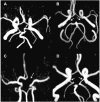
Fig. (2). Common Variants of circle of Willis.
Panel A. Complete circle of Willis. Panel B. Absent Posterior communicating arteries bilaterally (arrows). Panel C. Absent left A1 segment of anterior cerebral artery (arrow). Panel D bilateral fetal origin of posterior cerebral arteries (arrows), i.e. arising from the internal carotid arteries.
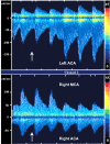
Fig. (3). Recruitment of collateral circulation during carotid artery endarterectomy, Transcranial Doppler study.
Inferior panel shows flow velocities in the proximal segment of the right middle cerebral artery (MCA) and right anterior cerebral artery (ACA). Two beats after clamping of the right internal carotid artery (arrow) there is a dramatic decrease in flow velocities in the ipsilateral middle cerebral artery and a less pronounced decrease in the ipsilateral anterior cerebral artery. The superior panel demonstrates increase in the flow velocity in the left anterior cerebral artery (negative deflection), contralateral to the clamped internal carotid artery, showing recruitment of collateral circulation.
Fig. (4). Transcranial Doppler Ultrasound demonstrating patterns of collateral circulation.
Panel A. Normal flow velocities and direction of flow in the internal (ICA), middle (MCA), and anterior cerebral arteries (ACA), and ophthalmic artery. Panels B and C. Example of recruitment of collateral circulation through the anterior cerebral artery (Panel B) and ophthalmic artery (Panel C) in a patient with right internal carotid occlusion below the level of the ophthalmic artery origin. Note that there is inversion of flow direction in the ACA (dotted arrow), compared to the normal side (arrow head). Panel C shows retrograde flow through the ophthalmic artery (solid arrow), demonstrating collateral circulation.
Fig. (5). Cerebral perfusion studies: Computed Tomography and Magnetic Resonance Imaging.
Panel A. Cerebral Blood Volume (CBV) demonstrates normal values. Panel B. Prolonged mean transit time (MTT) demonstrates hypoperfusion of the right hemisphere in the middle cerebral artery territory. Comparison of the CBV and MTT images reveals a mismatch, consistent with a region of ischemic penumbra. Panel C. Cerebral angiogram in the same patient showing collateral flow to the right anterior cerebral artery territory through the anterior communicating artery and leptomeningeal collaterals (arrow heads). Panels D, E and F. Example of diffusion weighted images (DWI) and perfusion weighted images (PWI) in a patient with carotid artery occlusion. Note small areas of restricted diffusion in Panel D (acute infarcts-bright signal) compared with a large area of decreased perfusion in Panel E (arrow), demonstrating a region of mismatch (ischemic penumbra). Panel F shows a three dimensional reconstruction of the carotid system based on CT angiography, demonstrating occlusion of the internal carotid artery at the origin (red arrow).
Fig. (6). Cerebral Angiography in a patient with right internal carotid artery occlusion.
Panels A and B. Leptomeningeal pial collateral channels (arrow heads) to the right anterior and middle cerebral artery territory (Panel A early arterial phase, Panel B late arterial phase). Panel C. Retrograde filling of intracranial internal carotid artery (arrow heads) through the ophthalmic artery (dotted arrow). Stenosis of the extracranial internal carotid artery in the proximal segment at the origin (black arrow) and occlusion in the distal extracranial segment (white solid arrow) are seen. Panel D. Collateral flow to the right anterior (solid arrow) and middle cerebral (dotted arrow) arteries through the anterior communicating artery (large solid arrow), in same patient with right internal carotid artery occlusion.
Fig. (7). Example of patient with carotid artery occlusion and clinical fluctuations related to hemodynamic factors; evaluation of collateral circulation and cerebral perfusion.
A 56-year-old right-handed man presented with confusion and left side weakness. He was found to have complete occlusion of the right internal carotid artery. Brain MRI demonstrated small infarctions in borderzone territories, located between anterior and middle cerebral artery, and between posterior and middle cerebral artery territories. Panel A demonstrates bright signal in the right hemisphere (Diffusion Weighted Imaging) and corresponding dark signal (Apparent Diffusion Coefficient, right of panel A) consistent with acute infarction. Magnetic Resonance Angiography (Panel B) and Computed Tomography Angiography of the neck and head (Panel F) confirmed occlusion of the proximal internal carotid artery after its origin (arrows). Panels B and E demonstrate lack of visualization of the right internal carotid artery intracranially as well as collateral flow through the anterior communicating artery suggested by visualization of the middle cerebral artery ipsilateral to the carotid occlusion. The patient presented severe worsening of left side weakness when anti hypertension medications were started, and developed left hemiplegia. A perfusion study (CTP) showed prolonged meant transit time (Panel C) and normal cerebral blood volume in the right middle cerebral artery territory (Panel D), consistent with a mismatch (ischemic penumbra). Repeat brain MRI showed no extension of the infarction (not shown). Collateral flow failure was suspected and blood pressure was elevated with intravenous fluids. The patient presented marked improvement and remained stable thereafter. He was discharged to rehabilitation, and at follow up 2 months later, had minimal left hemiparesis, was able to ambulate without supportive device and able to use fully his left hand. Modified Rankin Scale score was 1.

Similar articles

Cited by

References

    1. Barnett HJ, Taylor DW, Eliasziw M, et al. Benefit of carotid endarterectomy in patients with symptomatic moderate or severe stenosis. North american symptomatic carotid endarterectomy trial collaborators. N Engl J Med. 1998;339:1415–25. - PubMed
    1. Johnston SC, O'Meara ES, Manolio TA, et al. Cognitive impairment and decline are associated with carotid artery disease in patients without clinically evident cerebrovascular disease. Ann Intern Med. 2004;140:237–47. - PubMed
    1. van Oijen M, de Jong FJ, Witteman JC, et al. Atherosclerosis and risk for dementia. Ann Neurol. 2007;61:403–10. - PubMed
    1. Sacco RL. Clinical practice. Extracranial carotid stenosis. N Engl J Med. 2001;345:1113–8. - PubMed
    1. AbuRahma AF, Stone PA, Abu-Halimah S, Welch CA. Natural history of carotid artery occlusion contralateral to carotid endarterectomy. J Vasc Surg. 2006;44:62–66. - PubMed