. 2010 Oct;40(10):2891-901.
doi: 10.1002/eji.201040485.

Acquisition of regulatory function by human CD8(+) T cells treated with anti-CD3 antibody requires TNF

Affiliations

Acquisition of regulatory function by human CD8(+) T cells treated with anti-CD3 antibody requires TNF

Vitaly Ablamunits et al. Eur J Immunol. 2010 Oct.

Abstract

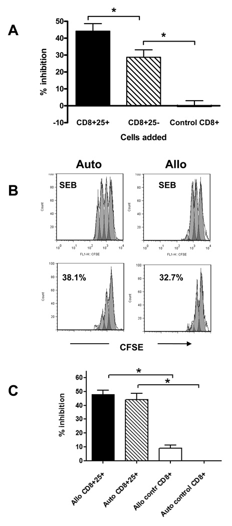
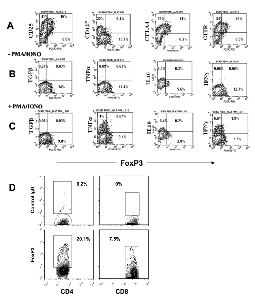
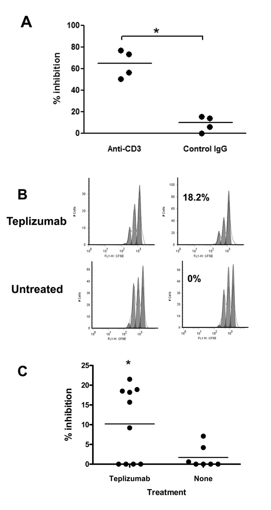
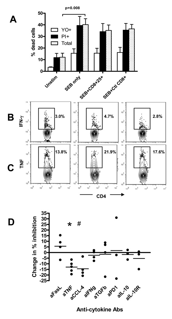
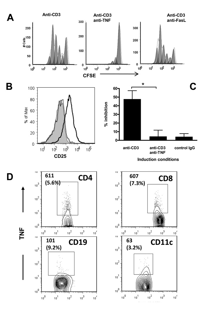
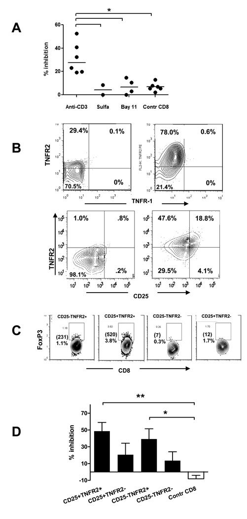
Anti-CD3 mAb can modulate graft rejection and attenuate autoimmune diseases but their mechanism(s) of action remain unclear. CD8(+) T cells with regulatory function are induced in vitro by Teplizumab, a humanized anti-CD3 antibody and inhibit responses of autologous and allogeneic T cells. They inhibit CD4(+) T-cell proliferation by mechanisms involving TNF and CCL4, and by blocking target cell entry into G2/M phase of cell cycle but neither kill them, nor compete for IL-2. CD8(+) Treg can be isolated from peripheral blood following treatment of patients with Type 1 diabetes with Teplizumab, but not from untreated patients. The induction of CD8(+) Treg by anti-CD3 mAb requires TNF and signaling through the NF-κB cascade. The CD8(+) Treg express CD25, glucocorticoid-induced TNF receptor family, CTLA-4, Foxp3, and TNFR2, and the combined expression of TNFR2 and CD25 identifies a potent subpopulation of CD8(+) Treg. These studies have identified a novel mechanism of immune regulation by anti-CD3 mAb and markers that may be used to track inducible CD8(+) Treg in settings such as chronic inflammation or immune therapy.


Conflict of interest statement

Conflicts of interests

The authors declare that they do not have conflicts of interests.

Figures

Figure 1
Figure 1. Inhibition of proliferative responses by CD8+ Tregs induced from PBMC from healthy donors
(A) PBMC from healthy control subjects were incubated with Teplizumab or control IgG for 5 days and CD8+ T cells were sorted on the basis of expression of CD25: CD8+CD25+ or CD8+CD25− cells from Teplizumab cultures or CD8+ cells from control IgG cultures. The cells were then incubated with autologous CD8-depleted PBMC and SEB for 72h, and inhibition of proliferation was calculated as described in Materials and Methods. Data are from 12 independent experiments (*P<0.01, ANOVA). (B) CD8+ 25+ cells were incubated with CD8-depleted autologous or allogeneic PBMC labeled with CFSE in the presence of SEB for 3 days and inhibition of proliferation measured. (a representative experiment of 3 is shown). (C) The pooled data from 3 independent experiments with 6 donors show a summary of the inhibition of anti-SEB responses by autologous or allogeneic CD8+CD25+ cells cultured with anti-CD3 or by CD8+ controls (*p<0.05 by ANOVA).
Figure 2
Figure 2. Phenotypic characterization of CD8+ T cells from healthy control subjects after 5 days of culture with anti-CD3 mAb
(A) PBMC were gated on CD8+ cells and stained for CD25, CD127, CTLA4, GITR, and FoxP3. A representative experiment of 4–8 independent experiments is shown. (B, C) PBMC cultured with anti-CD3 for 5 days were washed, rested in the absence of the mAb for three days, and then restimulated with PMA and ionomycin (C) or left untreated (B) for 6 hours in the presence of Golgi block. The cells were subsequently stained for intracellular cytokines and analyzed by flow cytometry. The plots show gated CD8+ populations with quadrant tool placed on the basis of isotype controls. The percentage of the gated cells is shown in each quadrant. A representative of 3 separate experiments with similar results is shown. (D) Comparison of relative numbers of FoxP3+ CD4+ (left) and CD8+ (right) cells 5 days after incubation with anti-CD3; a representative experiment out of 4 is shown. The percentage of cells within the indicated region (set based on isotype control) are shown.
Figure 3
Figure 3. Generation of iCD8+Tregs from patients with Type 1 diabetes in vitro and in vivo
(A) PBMC from patients with T1DM were cultured with anti-CD3 for 5 days, at which time, CD8+ cells were magnetically separated. The cells were incubated with allogeneic CD8-depleted and CFSE labeled cells plus SEB for 72h and inhibition of proliferation was calculated as described in Materials and Methods. Data from 4 individuals are shown (*P<0.05 compared to cells incubated with control IgG; paired t-test). (B, C) Induction of CD8+Tregs in vivo after administration of Teplizumab to T1DM patients. Proliferation of CD8-depleted CFSE-labeled responder cells in the presence of CD8+ T cells isolated from a representative drug-treated or control subject at the 1st and 2nd visits corresponding to before and after the drug treated subjects received anti-CD3 mAb(B). (C) Data from all 10 drug treated and 7 untreated patients tested. The percentage of inhibition was calculated as described in Materials and Methods (*p<0.05, paired t-test).
Figure 4
Figure 4. Analysis of potential mechanisms involved in inhibition by iCD8+Tregs generated from PBMCs of healthy donors
(A) CD8-depleted target cells were activated with SEB for three days in the presence or absence of CD8+CD25+ T cells from Teplizumab or CD8+ cells from control Ig cultures and apoptosis (YO+) and necrosis (PI+) were analyzed on gated CD4+ cells as described in Materials and Methods. Pooled data from five independent experiments are shown. (B) Addition of iCD8+Tregs does not inhibit cytokine production by target cells in response to SEB as judged by staining for intracellular IFNγ or TNF. Gated CD4+ cells are shown with a region placed on staining with control IgG. A representative of two similar experiments show a proportion of cytokine+ CD4+ cells cultured with SEB only (left panels), SEB+ iCD8+Tregs (middle panels), or SEB+control CD8+ T cells (right panels). (C) The effects of the indicated neutralizing antibodies on inhibition of proliferation by iCD8+Tregs are shown. The change in % inhibition was calculated from the percentage of dividing cells as: (% dividing cells with neutralizing antibody-% dividing cells with control Ig)/%dividing cells with control IgG. (#p=0.005,*p=0.032; by t-test compared to control Ig, n=5 for anti-TNF, n=4 for anti-CCL4, anti-FasL, anti-IFNg, and anti-TGFb; n=3 for anti-PD1, anti-IL-10, and anti-IL-10R).
Figure 5
Figure 5. Effects of TNF neutralization on the generation of iCD8+Tregs from PBMC of healthy control donors by anti-CD3 mAb in vitro
(A) CFSE-labeled PBMC were incubated with anti-CD3, with or without neutralizing anti-TNF or anti-FasL mAb for 5 days, and the proliferation and expression of CD25 on CD8+ cells was analyzed by FACS with a gate placed on CD8+ cells. A representative of three separate experiments is shown. (B) Up-regulation of CD25 on CD8+ T cells induced by anti-CD3 (bold histogram) on day 5 of culture is abrogated in the presence of anti-TNF (dotted histogram). A gate is placed on CD8+ T cells. The shaded peak shows CD25 expression on CD8+ cells incubated with normal human IgG. A representative of five separate experiments is shown. (C) Total CD8+ T cells isolated from cultures with Teplizumab with or without anti-TNF antibody were added to CD8-depleted PBMC labeled with CFSE and cultured with SEB. The inhibition of the target T cell proliferation was measured as described (*P=0.03 by ANOVA). Pooled data from 7 independent experiments are shown. (D) Analysis of the cell population responsible for TNF production after anti-CD3-induced activation by intracellular staining for TNF. Absolute numbers (and percent of the indicated subpopulation) of TNF+ cells counted during analysis of 30,000 PBMC with a gate placed on the lymphocyte subpopulation based on scatter. A representative of three independent experiments with similar results is shown. The region was set based on control IgG staining.
Figure 6
Figure 6. TNF signaling is needed to generate iCD8+Tregs from PBMC of healthy donors
(A) PBMC were cultured with anti-CD3 mAb with or without the inhibitors of NF-kB cascade Sulfasalazine or Bay 11–7082 for 5 days, total CD8+ cells were sorted, and added to autologous CD8-depleted, CFSE-labeled PBMC activated with SEB. The percent inhibiton of proliferation was calculated on Day 3 as described in Materials and Methods (*P<0.05 by ANOVA). (B) Normal human PBMC were incubated with anti-CD3 mAb for 5 days (right panels) or with control human IgG (left panels), and gated CD8+ cells were analyzed for TNFR-2 and TNFR1 expression (upper panels), or for TNFR-2 and CD25 expression (lower panels). Quadrants are built on the basis of isotype control IgG staining. A representative of three separate experiments is shown. (C) Normal human PBMC were incubated with anti-CD3 mAb for 5 days and stained for CD8, TNFR2, CD25, and FoxP3. The panels show CD8+ cells gated on the basis of CD25 and TNFR2 expression. The numbers in the upper left corner indicate the percentage and absolute numbers of CD8+Foxp3+ cells from 100,000 total lymphocytes collected. (D) PBMC incubated with anti-CD3 and CD8+ T cells sorted based on the expression of CD25 and TNFR-2 were tested for their ability to inhibit proliferation of target cells. (pooled data of 5 separate experiments is shown) (*P<0.05; **P<0.01; ANOVA).

Similar articles

Cited by

References

    1. Shou L, Schwartz SA, Good RA. Suppressor cell activity after concanavalin A treatment of lymphocytes from normal donors. J Exp Med. 1976;143:1100–1110. - PMC - PubMed
    1. Sampson D, Kauffman HM, Jr, Grotelueschen C, Metzig J. Suppressor activity of the human spleen and thymus. Surgery. 1976;79:393–397. - PubMed
    1. Hubert C, Delespesse G, Govaerts A. Concanavalin A-activated suppressor cells in normal human peripheral blood lymphocytes. Clin Exp Immunol. 1976;26:95–98. - PMC - PubMed
    1. Walker MR, Kasprowicz DJ, Gersuk VH, Benard A, Van Landeghen M, Buckner JH, Ziegler SF. Induction of FoxP3 and acquisition of T regulatory activity by stimulated human CD4+CD25− T cells. J Clin Invest. 2003;112:1437–1443. - PMC - PubMed
    1. Bettelli E, Oukka M, Kuchroo VK. T(H)-17 cells in the circle of immunity and autoimmunity. Nat Immunol. 2007;8:345–350. - PubMed

Publication types

MeSH terms