Comparative Study
. 2011 Feb;162(4):947-60.
doi: 10.1111/j.1476-5381.2010.01094.x.

Pharmacological characterization of six trkB antibodies reveals a novel class of functional agents for the study of the BDNF receptor

Affiliations
Comparative Study

Pharmacological characterization of six trkB antibodies reveals a novel class of functional agents for the study of the BDNF receptor

M Cazorla et al. Br J Pharmacol. 2011 Feb.

Abstract

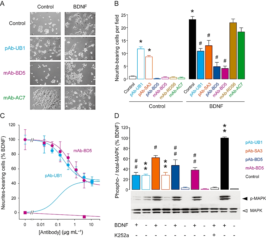
Background and purpose: By interacting with trkB receptors, brain-derived neurotrophic factor (BDNF) triggers various signalling pathways responsible for neurone survival, differentiation and modulation of synaptic transmission. Numerous reports have implicated BDNF and trkB in the pathogenesis of various central nervous system affections and in cancer, thus representing trkB as a promising therapeutic target. In this study, we used an antibody-based approach to search for trkB-selective functional reagents.

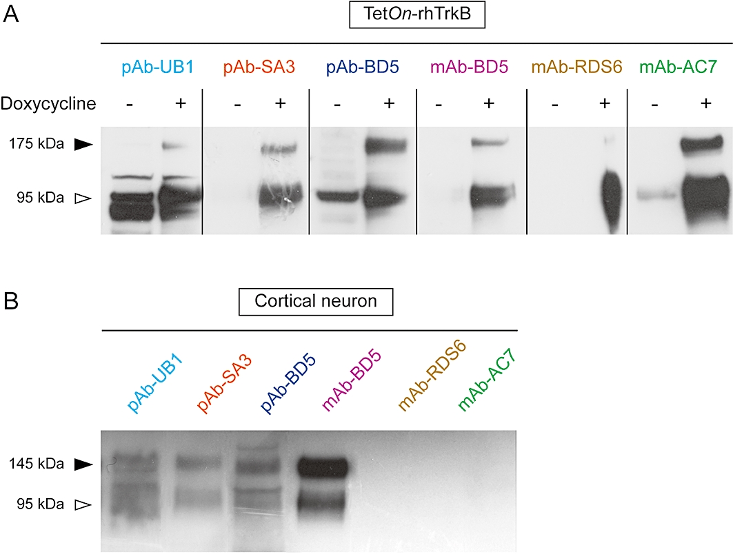
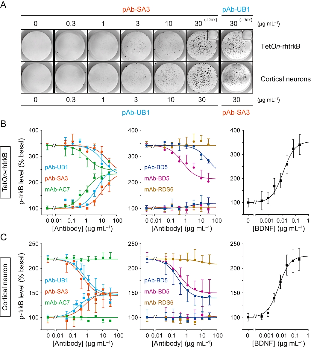
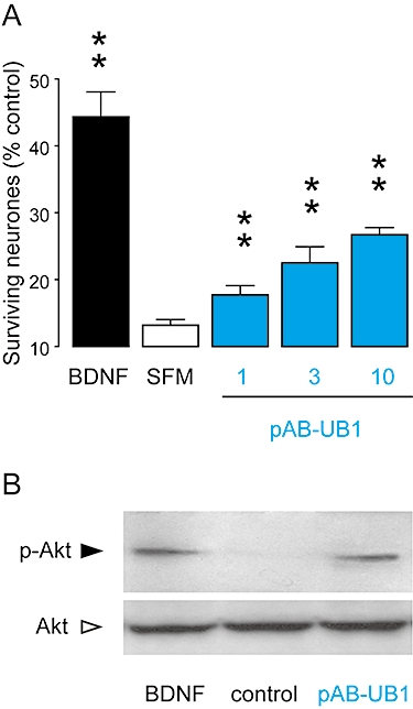
Experimental approach: Six commercially available polyclonal and monoclonal antibodies were tested on recombinant and native, human and rodent trkB receptors. Functional and pharmacological characterization was performed using a modified version of the KIRA-elisa method and radioligand binding studies. Western blot analyses and neurite outgrowth assays were carried out to determine the specificity and selectivity of antibody effects. The survival properties of one antibody were further assessed on cultured neurones in a serum-deprived paradigm.

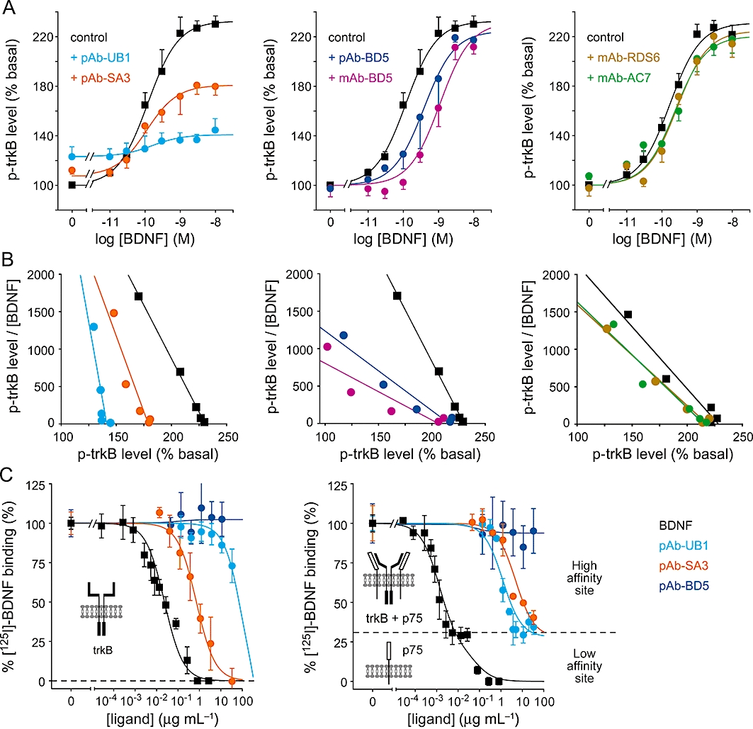
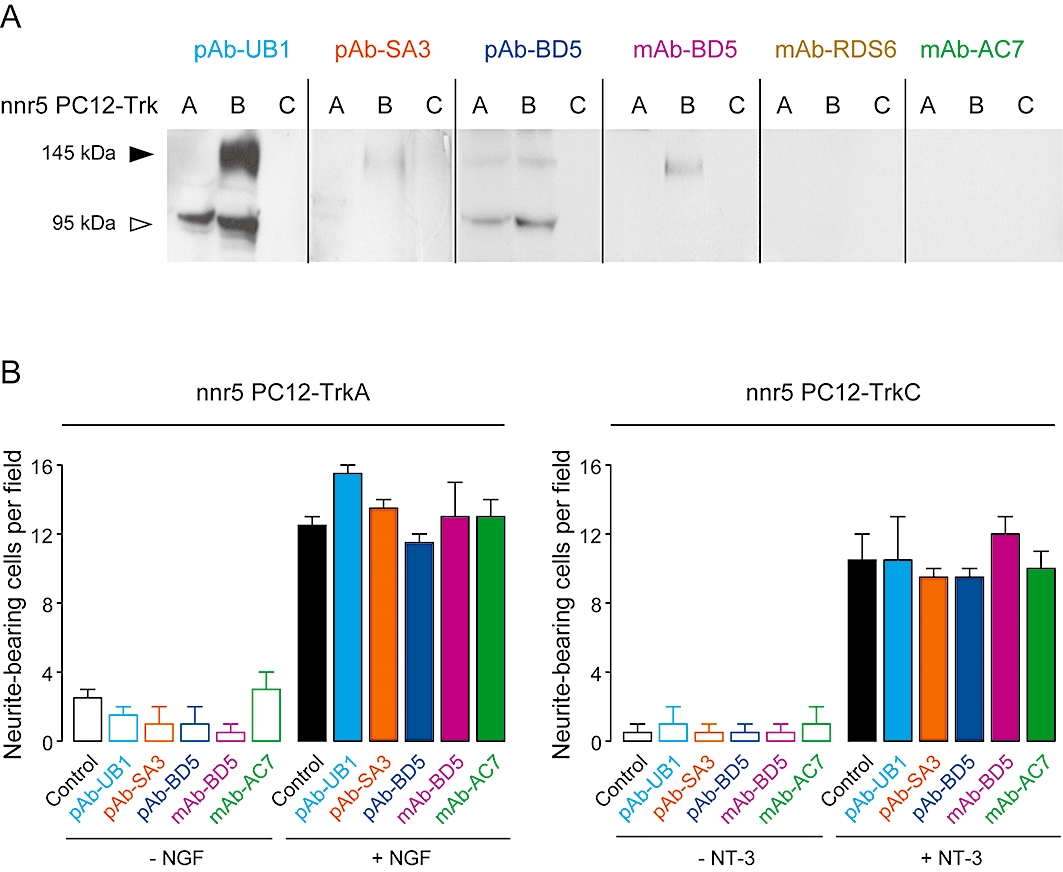
Key results: The functional trkB-selective antibodies showed distinct pharmacological profiles, ranging from partial agonists to antagonists, acting on trkB receptors through allosteric modulations. The same diversity of effects was observed on the mitogen-activated protein kinase signalling pathway downstream of trkB and on the subsequent neurite outgrowth. One antibody with partial agonist activity demonstrated cell survival properties by activating the Akt pathway. Finally, these antibodies were functionally validated as true trkB-selective ligands because they failed activating trkA or trkC, and contrary to BDNF, none of them bind to p75(NTR).

Conclusions and implications: These trkB-selective antibodies represent a novel class of pharmacological tools to explore the pathophysiological roles of trkB and its potential therapeutic relevance for the treatment of various disorders.


Figures

Figure 1
Figure 1
Rodent and human cross-reactive responses for pAbs and mAbs. Antibodies (1 µg·mL−1) were subjected to Western blot analysis to examine their binding properties to (A) human and (B) mouse trkB, in TetOn-rhtrkB treated with or without doxycycline, and neuronal cells respectively. The trkB signal elicits two bands, corresponding to the full length (145 kDa) and truncated (95 kDa) form of the receptor. In TetOn-rhtrkB cells, trkB is C-terminally fused to the fluorescent protein ECFP, resulting in the detection of two bands of approximately 175 kDa (trkB + ECFP) and 95 kDa (truncated trkB). pAbs, polyclonal antibodies; mAbs, monoclonal antibodies; TetOn, tetracycline-responsive; ECFP, enhanced cyan fluorescent protein.
Figure 2
Figure 2
Distinct functional modulations of trkB activity by pAbs and mAbs in both recombinant and native systems. The phosphorylation levels of trkB were quantified by KIRA-elisa assays after treatment with BDNF and antibodies, alone or in combination. (A) Representative fluorescent photomicrographs of TetOn-rhtrkB cells and cortical neurones after treatment with increasing concentrations of pAb-SA3 and pAb-UB1 as for KIRA-elisa assays. Insets show signal obtained with 30 µg·mL−1 of pAb-UB1 or pAb-SA3 in non-induced TetOn-rhtrkB cells (-Dox). (B,C) Increasing concentrations of pAbs and mAbs in the presence (circles) or absence (squares) of BDNF (B, TetOn-rhtrkB, 1 nM; C, neurones, 0.4 nM). BDNF concentration-response curves are shown for comparison. Absorbance read at 450 nm was normalized as percentage of basal trkB activity. Data are mean ± SEM of values obtained in triplicate in three experiments. pAbs, polyclonal antibodies; mAbs, monoclonal antibodies; TetOn, tetracycline-responsive; BDNF, brain-derived neurotrophic factor.
Figure 3
Figure 3
Mechanism of action of pAbs and mAbs. (A) BDNF concentration-response curves in the absence or presence of antibodies (10 µg·mL−1) were obtained in neurones. The addition of pAb-UB1 and pAb-SA3 (left panel) resulted in a non-competitive partial agonism and did not significantly change BDNF EC50. pAb-BD5 and mAb-BD5 (middle panel) elicited a competitive effect, as demonstrated by the significant rightward shift of the BDNF curve. mAb-RDS6 and mAb-AC7 (right panel) were inactive in neurones. (B) Eadie–Hofstee plots of the data obtained in (A) confirmed the non-competitive actions of pAb-UB1 and pAb-SA3 (parallel lines, left panel), the competitive effect elicited by pAb-BD5 and mAb-BD5 (convergent lines, middle panel) and the lack of effect of mAb-RDS6 and mAb-AC7 (right panel). Values are expressed as percentage of basal trkB activity. Data are mean ± SEM of 3–4 experiments performed in triplicate, except for Eadie–Hofstee plots where data are means. (C) Effect of pAbs on the binding of [125I]-BDNF in TetOn-rhtrkB cells (left panel) and in neurones (right panel). Insets show the conformations of the receptors supposed to be responsible for the different BDNF binding sites. Data are mean ± SEM of 2–4 experiments performed in triplicate. pAbs, polyclonal antibodies; mAbs, monoclonal antibodies; BDNF, brain-derived neurotrophic factor; TetOn, tetracycline-responsive.
Figure 4
Figure 4
Binding and functional selectivity of pAbs and mAbs for TrkB but not for TrkA or TrkC receptors. (A) Antibodies (10 µg·mL−1) were subjected to Western blot analysis to assess their selectivity with regard to trkA and trkC using nnr5 PC12-trkA and -trkC cells. A representative immunoblot is presented. (B) Quantitative analysis of NGF- (2 nM) and NT3 (10 nM) -induced neurite outgrowth in the presence of 10 µg·mL−1 of antibodies in nnr5 PC12-trkA and -trkC cells respectively. Data are mean ± SEM of results from two experiments. pAbs, polyclonal antibodies; mAbs, monoclonal antibodies; NGF, nerve growth factor.
Figure 5
Figure 5
Biological effects of pAbs and mAbs on neurite outgrowth and trkB-dependent MAPK signalling activation. (A) Representative photomicrographs of nnr5 PC12-trkB cells treated for 48 h with 10 µg·mL−1 antibodies (partial agonist effects illustrated by pAb-UB1; antagonist effects illustrated by mAb-BD5; inactive antibody illustrated by mAb-AC7) and 1 nM BDNF, as indicated. (B) Quantitative analysis of BDNF-induced neurite outgrowth in the presence of antibodies. (C) Concentration-response curves of partial agonist pAb-UB1 and antagonist mAb-BD5 obtained in the presence or absence of 1 nM BDNF were performed as in (B). Values are expressed as a percentage of the maximal BDNF response (100%) after subtraction of counting obtained in controls (0%). Data are mean ± SEM of results from two experiments. (D) Representative Western blot analysis of total and phosphorylated MAPK in nnr5 PC12-trkB cells after treatment with BDNF (1 nM), antibodies (10 µg·mL−1) or K252a (100 nM), alone or in combination. Densitometric quantification of MAPK phosphorylation relative to total MAPK is shown and expressed as a percentage of BDNF response after subtraction of background values. Data are mean ± SEM of values obtained in triplicate in two independent experiments; **P < 0.01, compared with basal condition; ##P < 0.01, compared with BDNF; *P < 0.05; #P < 0.05. pAbs, polyclonal antibodies; mAbs, monoclonal antibodies; MAPK, mitogen-activated protein kinase; BDNF, brain-derived neurotrophic factor.
Figure 6
Figure 6
Biological effects of pAbs and mAbs on neurone survival. (A) Increasing concentrations of pAb-UB1 or BDNF (1 nM) were added to cultured neurones for 3 days after serum deprivation. Cell viability data are expressed as percentage of total living cells in the presence of complete culture medium and are mean ± SEM of values obtained in triplicate in three independent experiments; **P < 0.01, compared with serum-deprived condition. (B) Representative immunoblotting analysis of total and phosphorylated Akt in cortical neurones after treatment with BDNF (1 nM) or pAb-UB1 (10 µg·mL−1). pAbs, polyclonal antibodies; mAbs, monoclonal antibodies; BDNF, brain-derived neurotrophic factor; SFM, serum-free medium.

References

    1. Alexander SPH, Mathie A, Peters JA. Guide to Receptors and Channels (GRAC) Br J Pharmacol. 2009;158(Suppl 1):S1–S254. 4th edn. - PMC - PubMed
    1. Banfield MJ, Naylor RL, Robertson AG, Allen SJ, Dawbarn D, Brady RL. Specificity in Trk receptor: neurotrophin interactions: the crystal structure of TrkB-d5 in complex with neurotrophin-4/5. Structure (Camb) 2001;9:1191–1199. - PubMed
    1. Benedetti M, Levi A, Chao MV. Differential expression of nerve growth factor receptors leads to altered binding affinity and neurotrophin responsiveness. Proc Natl Acad Sci USA. 1993;90:7859–7863. - PMC - PubMed
    1. Cattaneo A, Capsoni S, Margotti E, Righi M, Kontsekova E, Pavlik P, et al. Functional blockade of tyrosine kinase A in the rat basal forebrain by a novel antagonistic anti-receptor monoclonal antibody. J Neurosci. 1999;19:9687–9697. - PMC - PubMed
    1. Cazorla M, Jouvenceau A, Rose C, Guilloux JP, Pilon C, Dranovsky A, et al. Cyclotraxin-B, the first highly potent and selective TrkB inhibitor, has anxiolytic properties in mice. PLoS One. 2010;5:e9777. - PMC - PubMed

Publication types

MeSH terms