. 2011 Feb;132(2):217-25.
doi: 10.1111/j.1365-2567.2010.03356.x. Epub 2010 Oct 13.

Differences in T-helper polarizing capability between human monocyte-derived dendritic cells and monocyte-derived Langerhans'-like cells

Affiliations

Differences in T-helper polarizing capability between human monocyte-derived dendritic cells and monocyte-derived Langerhans'-like cells

Ivan Rajkovic et al. Immunology. 2011 Feb.

Abstract

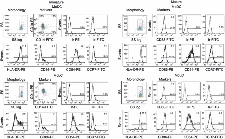
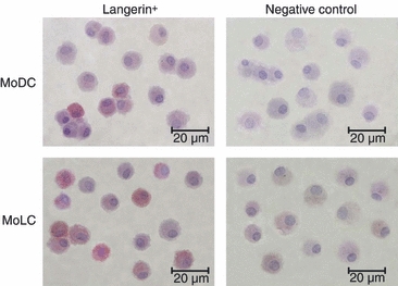
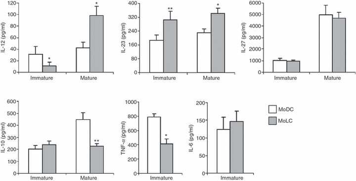
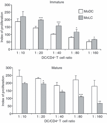
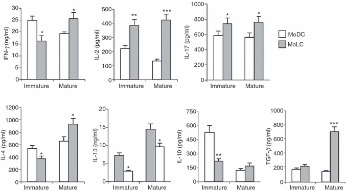
Langerhans' cells (LCs) represent a specific subset of dendritic cells (DCs) which are important for detecting and processing pathogens that penetrate the skin and epithelial barriers. The aim of our study was to explain what makes their in vitro counterparts - monocyte-derived Langerhans'-like cells (MoLCs) - unique compared with monocyte-derived dendritic cells (MoDCs). Immature MoDCs were generated by incubating peripheral blood monocytes with granulocyte-macrophage colony-stimulating factor (GM-CSF) and interleukin (IL)-4. The addition of transforming growth factor-β (TGF-β) to this cytokine cocktail resulted in the generation of MoLCs. MoLCs showed a lower expression of CD83, CD86, HLA-DR and CCR7 compared with MoDCs, regardless of their maturational status. Both immature and mature MoLCs secreted higher quantities of IL-23 compared with MoDCs and this finding correlated with a higher secretion of IL-17 in co-culture of MoLCs with allogeneic CD4(+) T cells. Mature MoLCs, which produced higher levels of IL-12 and lower levels of IL-10 compared with mature MoDCs, were more potent at inducing interferon-γ (IFN-γ) production by CD4(+) T cells in the co-culture system. In conclusion, the finding that mature MoLCs stimulate stronger T-helper 1 and T-helper 17 immune responses than mature MoDCs, makes them better candidates for use in the preparation of anti-tumour DC vaccines.


Figures

Figure 1
Figure 1
Phenotypic characterization of monocyte-derived dendritic cells (MoDCs) and monocyte-derived Langerhans’ like cells (MoLCs), as determined by flow cytometry. MoDCs were generated from peripheral blood monocytes after incubation with granulocyte–macrophage colony-stimulating factor (GM-CSF) and interleukin-4 (IL-4). MoLCs were generated under the same conditions but with the addition of transforming growth factor- β (TGF-β) to the cytokine cocktail. Maturation of these DC subsets was induced with a cocktail of proinflammatory mediators, as described in the Materials and methods. The numbers in the upper right corners of single histograms represent mean values of fluorescence intensity within the gated populations [marked on forward scatter (FS) : side scatter (SS) log profiles]. The results are representative of one donor, out of five different experiments. Ir-PE and Ir-FITC represent non-specific (background) fluorescence using irrelevant (Ir) mouse monoclonal antibodies (mAbs) conjugated to the corresponding fluorochrome [i.e. phycoerythrin (PE) and fluorescein isothiocyanate (FITC)], which do not react with human cells.
Figure 2
Figure 2
Phenotypic characterization of monocyte-derived dendritic cells (MoDCs) and monocyte-derived Langerhans’ like cells (MoLCs), as determined by immunocytochemistry. Cytospin preparations of immature dendritic cell (iDC) samples were stained with anti-human Langerin IgG, as described in the Materials and methods. At least 500 cells were analyzed on each cytospin using a light microscope and the percentage of positive cells was determined. Original magnification, × 600; the bar represents 20 μm. The slides with the omitted primary antibody served as negative controls.
Figure 3
Figure 3
Production of cytokines in monocyte-derived dendritic cells (MoDCs) and monocyte-derived Langerhans’ like cells (MoLCs) culture supernatants. The supernatants of both immature and mature MoDCs and MoLCs were collected after 6 days of culture, and the concentrations of cytokines (in pg/ml) were determined using enzyme-linked immunosorbent assays (ELISAs). Values are given as mean ± standard deviation (SD) of three different experiments. *P < 0·05, **P< 0·01 compared with MoDCs. IL, interleukin; TNF-α, tumour necrosis factor-α.
Figure 4
Figure 4
Allostimulatory activity of monocyte-derived dendritic cells (MoDCs) and monocyte-derived Langerhans’ like cells (MoLCs). Immature and mature MoDCs and MoLCs were co-cultured with allogeneic CD4+ T cells at different ratios. After 5 days, the cultures were pulsed with [3H]thymidine for the last 18 hr and the radioactivity was measured as described in the Materials and methods. Values are given as mean ± standard deviation (SD) of triplicate samples from one representative experiment out of three different experiments showing similar results. The basal counts per minute (c.p.m.) in CD4+ T-cell cultures, alone, was 402 ± 53 c.p.m. The basal c.p.m. in DC cultures alone, independently of number and type of DCs, was between 62 and 102 c.p.m. (similar to the background radioactivity). The proliferation index (PI) was calculated as follows: PI = c.p.m. (DC/CD4+ T-cell co-culture)/[c.p.m. (CD4+ T-cell culture alone) + c.p.m. (DC culture alone)]. *P < 0·05, **P < 0·01, ***P < 0·005 compared with MoDCs.
Figure 5
Figure 5
Production of cytokines by allogeneic CD4+ T cells in co-culture with immature or mature monocyte-derived dendritic cells (MoDCs) or monocyte-derived Langerhans’ like cells (MoLCs). The amounts of cytokines in both co-cultures (MoDCs/CD4+ T cells and MoLCs/CD4+ T cells) were measured using enzyme-linked immunosorbent assays (ELISAs) or FlowCytomix Human Th1/Th2 11plex, as described in the Materials and methods. Values are given as mean ± standard deviation (SD) of three independent experiments. *P < 0·05, **P < 0·01, ***P < 0·005 compared with MoDCs. IFN-γ, interferon-γ; IL, interleukin; TNF-α, tumour necrosis factor-α.

References

    1. Riedl E, Stockl J, Majdic O, Scheinecker C, Knapp W, Strobl H. Ligation of E-cadherin on in vitro-generated immature Langerhans-type dendritic cells inhibits their maturation. Blood. 2000;96:4276–84. - PubMed
    1. Merad M, Manz MG. Dendritic cell homeostasis. Blood. 2009;113:3418–27. - PMC - PubMed
    1. Banchereau J, Briere F, Caux C, et al. Immunobiology of dendritic cells. Annu Rev Immunol. 2000;18:767–811. - PubMed
    1. Poulin LF, Henri S, de Bovis B, Devilard E, Kissenpfennig A, Malissen B. The dermis contains langerin+ dendritic cells that develop and function independently of epidermal Langerhans cells. J Exp Med. 2007;204:3119–31. - PMC - PubMed
    1. Geissmann F, Dieu-Nosjean MC, Dezutter C, et al. Accumulation of immature Langerhans cells in human lymph nodes draining chronically inflamed skin. J Exp Med. 2002;196:417–30. - PMC - PubMed

Publication types