

Mobilization of plasma cells in healthy individuals treated with granulocyte colony-stimulating factor for haematopoietic stem cell collection
- PMID: 21039470
- PMCID: PMC3050449
- DOI: 10.1111/j.1365-2567.2010.03361.x
Mobilization of plasma cells in healthy individuals treated with granulocyte colony-stimulating factor for haematopoietic stem cell collection
Abstract
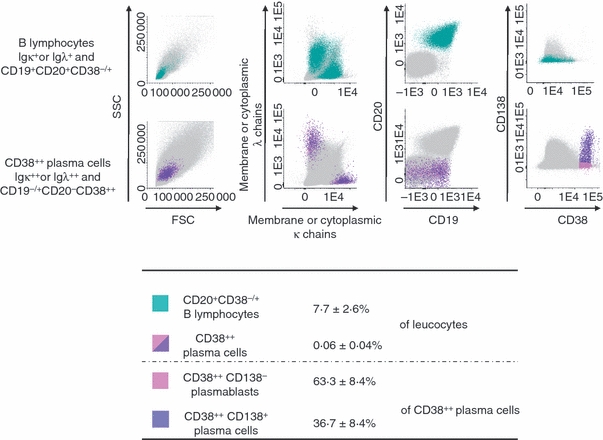
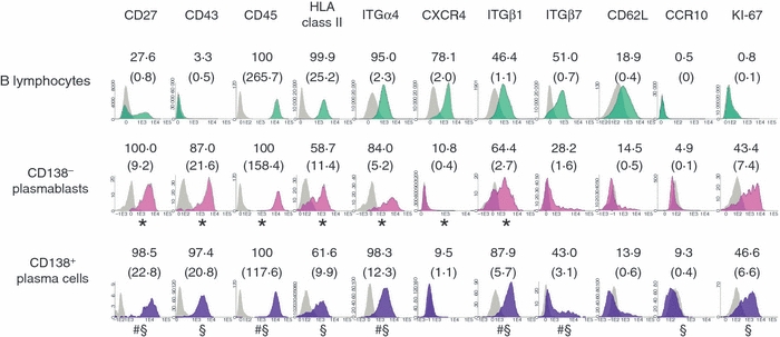
In mice, the plasma cell (PC) niche in the bone marrow is close to the haematopoietic stem cell (HSC) niche. We investigated whether PCs can be mobilized into the peripheral blood (PB) in healthy donors receiving granulocyte colony-stimulating factor (G-CSF) for the induction of HSC mobilization into the PB. G-CSF increased the count of circulating PCs 6-fold, that of circulating B lymphocytes 4-fold and that of circulating HSCs 44-fold. Mobilized circulating PCs comprised CD138(-) (62·2%) and CD138(+) (37·8%) PCs, the latter being more mature based on increased CD27, CD38 and cytoplasmic immunoglobulin expression. Mobilized PCs had a phenotype close to that of steady-state PB PCs or in vitro generated PCs, but they expressed L-selectin only weakly. Finally, a median value of 0·4 × 10(6) /kg donor PCs - one-thirtieth of the overall PC count in a healthy adult - was grafted into patients, which could contribute to immune memory recovery.
Figures
Similar articles
-
Functional analysis of human hematopoietic repopulating cells mobilized with granulocyte colony-stimulating factor alone versus granulocyte colony-stimulating factor in combination with stem cell factor.Blood. 2002 Aug 1;100(3):869-78. doi: 10.1182/blood.v100.3.869. Blood. 2002. PMID: 12130497
-
Platelet-derived circulating soluble P-selectin is sufficient to induce hematopoietic stem cell mobilization.Stem Cell Res Ther. 2023 Oct 20;14(1):300. doi: 10.1186/s13287-023-03527-w. Stem Cell Res Ther. 2023. PMID: 37864264 Free PMC article.
-
Effect of stem cell mobilization with cyclophosphamide plus granulocyte colony-stimulating factor on morphology of haematopoietic organs in mice.Cell Prolif. 2005 Feb;38(1):47-61. doi: 10.1111/j.1365-2184.2005.00329.x. Cell Prolif. 2005. PMID: 15679866 Free PMC article.
-
Granulocyte colony-stimulating factor versus granulocyte-macrophage colony-stimulating factor for collection of peripheral blood progenitor cells from healthy donors.Curr Opin Hematol. 2000 May;7(3):150-5. doi: 10.1097/00062752-200005000-00004. Curr Opin Hematol. 2000. PMID: 10786651 Review.
-
Mobilizing stem cells from normal donors: is it possible to improve upon G-CSF?Bone Marrow Transplant. 2007 May;39(10):577-88. doi: 10.1038/sj.bmt.1705616. Epub 2007 Mar 19. Bone Marrow Transplant. 2007. PMID: 17369869 Review.
Cited by
-
Fingerprints of CD8+ T cells on human pre-plasma and memory B cells.PLoS One. 2018 Dec 12;13(12):e0208187. doi: 10.1371/journal.pone.0208187. eCollection 2018. PLoS One. 2018. PMID: 30540814 Free PMC article.
-
Exercise as an Adjuvant Therapy for Hematopoietic Stem Cell Mobilization.Stem Cells Int. 2016;2016:7131359. doi: 10.1155/2016/7131359. Epub 2016 Mar 31. Stem Cells Int. 2016. PMID: 27123008 Free PMC article. Review.
-
Niches for Hematopoietic Stem Cells and Their Progeny.Immunity. 2018 Apr 17;48(4):632-648. doi: 10.1016/j.immuni.2018.03.024. Immunity. 2018. PMID: 29669248 Free PMC article. Review.
-
Application of mass cytometry to characterize hematopoietic stem cells in apheresis products of patients with hematological malignancies.Hematol Transfus Cell Ther. 2024 Dec;46 Suppl 6(Suppl 6):S59-S70. doi: 10.1016/j.htct.2023.10.008. Epub 2023 Dec 29. Hematol Transfus Cell Ther. 2024. PMID: 38177056 Free PMC article.
-
Residual malignant and normal plasma cells shortly after high dose melphalan and stem cell transplantation. Highlight of a putative therapeutic window in Multiple Myeloma?Oncotarget. 2012 Nov;3(11):1335-47. doi: 10.18632/oncotarget.650. Oncotarget. 2012. PMID: 23154454 Free PMC article.
References
-
- Radbruch A, Muehlinghaus G, Luger EO, Inamine A, Smith KG, Dorner T, Hiepe F. Competence and competition: the challenge of becoming a long-lived plasma cell. Nat Rev Immunol. 2006;6:741–50. - PubMed
-
- Tarlinton D, Radbruch A, Hiepe F, Dorner T. Plasma cell differentiation and survival. Curr Opin Immunol. 2008;20:162–9. - PubMed
-
- DiLillo DJ, Hamaguchi Y, Ueda Y, Yang K, Uchida J, Haas KM, Kelsoe G, Tedder TF. Maintenance of long-lived plasma cells and serological memory despite mature and memory B cell depletion during CD20 immunotherapy in mice. J Immunol. 2008;180:361–71. - PubMed
-
- Odendahl M, Mei H, Hoyer BF, et al. Generation of migratory antigen-specific plasma blasts and mobilization of resident plasma cells in a secondary immune response. Blood. 2005;105:1614–21. - PubMed
-
- Tokoyoda K, Egawa T, Sugiyama T, Choi BI, Nagasawa T. Cellular niches controlling B lymphocyte behavior within bone marrow during development. Immunity. 2004;20:707–18. - PubMed
Publication types
MeSH terms
Substances
LinkOut - more resources
Full Text Sources
Medical
Research Materials
