. 2010 Nov;32(9):1497-506.
doi: 10.1111/j.1460-9568.2010.07411.x. Epub 2010 Oct 12.

Neuropeptide-mediated calcium signaling in the suprachiasmatic nucleus network

Affiliations

Neuropeptide-mediated calcium signaling in the suprachiasmatic nucleus network

Robert P Irwin et al. Eur J Neurosci. 2010 Nov.

Abstract

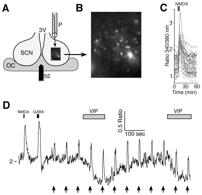
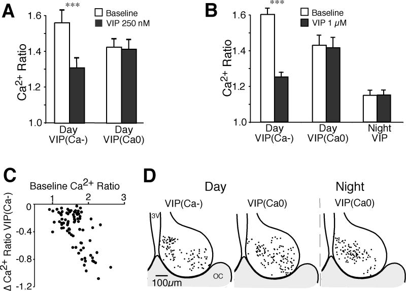
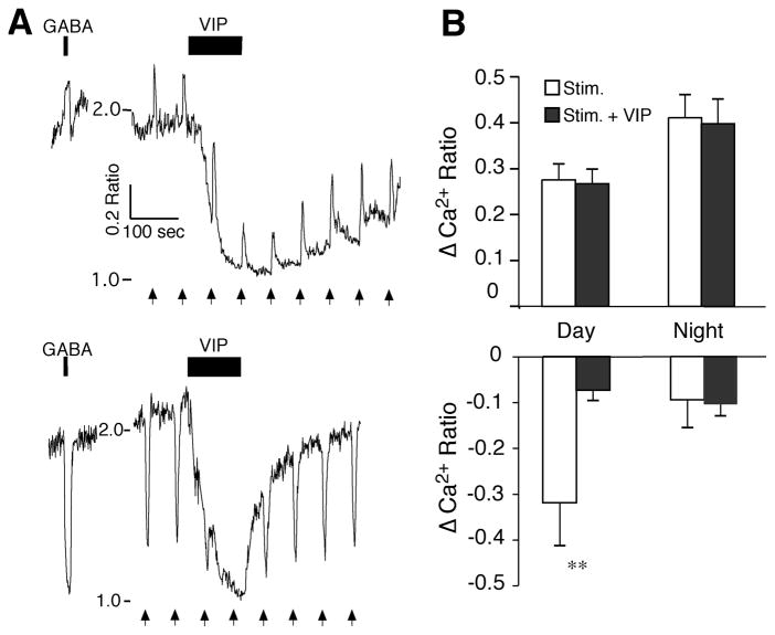
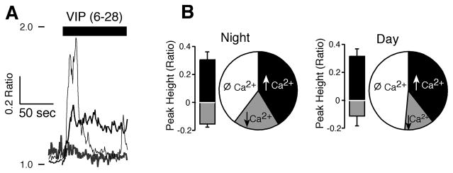
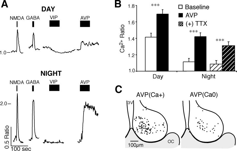
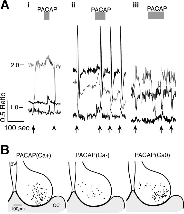
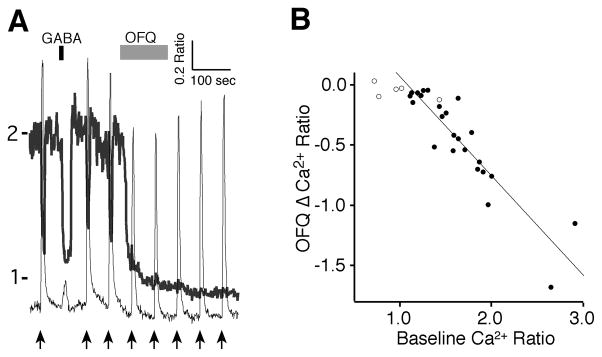
Neuroactive peptides and the intracellular calcium concentration ([Ca(2+) ](i) ) play important roles in light-induced modulation of gene expression in the suprachiasmatic nucleus (SCN) neurons that ultimately control behavioral rhythms. Vasoactive intestinal peptide (VIP) and arginine vasopressin (AVP) are expressed rhythmically within populations of SCN neurons. Pituitary adenylate cyclase-activating peptide (PACAP) is released from retinohypothalamic tract (RHT) terminals synapsing on SCN neurons. Nociceptin/orphanin FQ (OFQ) receptors are functionally expressed in the SCN. We examined the role of several neuropeptides on Ca(2+) signaling, simultaneously imaging multiple neurons within the SCN neural network. VIP reduced the [Ca(2+) ](i) in populations of SCN neurons during the day, but had little effect at night. Stimulation of the RHT at frequencies that simulate light input signaling evoked transient [Ca(2+) ](i) elevations that were not altered by VIP. AVP elevated the [Ca(2+) ](i) during both the day and night, PACAP produced variable responses, and OFQ induced a reduction in the [Ca(2+) ](i) similar to VIP. During the day, VIP lowered the [Ca(2+) ](i) to near nighttime levels, while AVP elevated [Ca(2+) ](i) during both the day and night, suggesting that the VIP effects on [Ca(2+) ](i) were dependent, and the AVP effects independent of the action potential firing activity state of the neuron. We hypothesize that VIP and AVP regulate, at least in part, Ca(2+) homeostasis in SCN neurons and may be a major point of regulation for SCN neuronal synchronization.


Figures

Figure 1
Figure 1. Measurement of intracellular Ca2+ simultaneously in multiple SCN neurons
A. Schematic illustration of the recording setup showing a coronal hypothalamic slice containing the SCN and third ventricle (3V) with a bipolar stimulating electrode (SE) used to stimulate the RHT positioned in the optic chiasm (OC) and a perfusion pipette (P) positioned to flow solutions over the SCN. B. High power (40X objective) fluorescent image of fura-2 loaded SCN neurons. C. Example showing NMDA (200 μM, 5 sec) inducing a transient increase in the Ca2+ ratio (340nm/380nm) in SCN neurons during the day (ZT = 6 hours). D. Example showing changes in the Ca2+ ratio of a SCN neuron with responses to NMDA (200 μM, 5 sec), GABA (200 μM, 10 sec), RHT stimulation (arrows; 100 pulses, 200 μs at 20 Hz) and VIP (1 μM) application.
Figure 2
Figure 2. VIP induces a reduction of baseline [Ca2+]i during the day but not the night
A. Comparison of the mean Ca2+ ratio of SCN neurons at baseline and following application of VIP (250 nM) during the day (n = 96). VIP (250 nM) induced a significant reduction from baseline in SCN neurons showing a VIP(Ca−) response (n = 26, t = 7.24, *** p < 0.0001), while most neurons had little or no response (VIP(Ca0), n = 61, t = 1.15, p = 0.25), and only 9 neurons had a Ca2+ increase (data not shown). B. Comparison of the mean Ca2+ ratio of SCN neurons at baseline and following application of VIP (1 μM) during the day (n = 152) and night (n = 121). During the day, VIP (1 μM significantly reduced the baseline Ca2+ ratio (mean ± SEM) in VIP(Ca−) neurons (n = 82, t = 11.9, *** p < 0.0001), but not in the VIP(Ca0) group (n = 59, t = 1.71, p = 0.093), while only 11 neurons showed a Ca2+ increase (data not shown). During the night, VIP reduced the Ca2+ ratio in 7% of neurons. The data shown is the mean ± SEM of all nighttime neurons (n = 121 neurons, t = 0.114, p = 0.91). Note the baseline Ca2+ ratios were higher during the day than the night, and that VIP reduced the Ca2+ ratios of VIP(Ca−) responding neurons to near nighttime levels. C. Plot of the baseline Ca2+ ratio versus the VIP (1 μM)-induced change in the Ca2+ ratio in VIP(Ca−) neurons during the day. Note that larger reductions in Ca2+ occur in neurons with higher baseline Ca2+ ratios (R2 = 0.41). D. Regional location of VIP-treated neurons during the day and night. The position for each VIP(Ca−) and VIP(Ca0) neuron was superimposed on a representative drawing of the SCN with the third ventricle (3V) on the left and optic chiasm (OC) at the bottom. Neither VIP(Ca−) or VIP(Ca0) neurons demonstrated a clear regional expression pattern.
Figure 3
Figure 3. VIP modulation of RHT signaling in the SCN network
A. The postsynaptic Ca2+ ratio in two SCN neurons with similar reductions in response to VIP (1 μM), but opposing responses to both GABA (200 μM, 10 sec) and stimulation of the RHT (arrows, 100 pulses, 200 μs at 20 Hz). B. VIP (1 μM) had little or no effect on RHT-evoked elevations of Ca2+ (top) during the day (n = 36 neurons, t = 0.43, p = 0.67) and night (n = 22, t = 0.39, p = 0.70), but attenuated RHT-evoked Ca2+ reductions during the day (n = 10, t = 2.82, ** p < 0.020), but not the night (n = 3, t = 1.06, p = 0.40). Data is the mean ± SEM of maximum (Ca2+ elevations) and minimum (Ca2+ reductions) responses to RHT-evoked changes in the Ca2+ ratio before and during VIP (1 μM) application. Note that since VIP reduced the baseline Ca2+, the magnitudes of RHT-evoked Ca2+ reductions were also reduced, while at night the baseline Ca2+ ratio was low with few RHT-evoked Ca2+ transient reductions.
Figure 4
Figure 4. The VIP antagonist VIP(6-28) modified [Ca2+]i in SCN neurons at night
A. Example showing three SCN neurons during the night with different responses to application of VIP(6-28) (2 μM), a transient rise, a prolonged elevation and a reduction in the Ca2+ ratio. B. Pie chart of VIP(6-28) data showing the proportion of neurons with elevations (↑, n = 22) reductions (↓, n = 10) and no responses (Ø, n = 21) in Ca2+. The bars represent the mean ± SEM of the maximal (black) and minimum (gray) responses (0–30 sec) of neurons showing VIP(6–28) (2–4 μM) evoked Ca2+ changes.
Figure 5
Figure 5. AVP elevates the [Ca2+]i of SCN neurons during the day and night
A. Example showing SCN neurons responding to AVP during the day and night. Tracings show treatments with NMDA (200 μM), GABA (200 μM), VIP (1 μM) and AVP (1 μM). Note that VIP lowered the Ca2+ ratio during the day but not at night, while AVP elevated the Ca2+ ratio [AVP(Ca+)] during both the day and night. B. Bar graph of AVP(Ca+) responding neurons showing the mean ± SEM Ca2+ ratio before and during AVP application during the day (n = 53, t = 8.25, *** p < 0.001) and night (n = 55, t = 8.44, *** p < 0.001), and during night with TTX (ANOVA, F3,54 = 28.6, p < 0.001, Bonferroni adjusted p = 0.48 [night-Baseline] and p = 0.09 [night-AVP]). C. Regional location of AVP-treated neurons (day and night combined). The position for each AVP(Ca+) and non-responding [AVP(Ca0)] neuron was superimposed on a representative drawing of the SCN with the third ventricle (3V) on the left and optic chiasm (OC) at the bottom. AVP(Ca+)-responding neurons appeared to be located primarily in the dorsomedial region.
Figure 6
Figure 6. The Effects of PACAP on [Ca2+]i in SCN neurons
A. Examples of the variety of Ca2+ responses observed with PACAP (1–38) application onto SCN neurons during the day. i & ii) PACAP (250 nM) evoked transient Ca2+ elevations without altering the RHT stimulation-induced transients (arrows, 100 pulses at 20 Hz). iii) PACAP (500 nM) evoked a Ca2+ reduction in one neuron and an elevation in another. B. Regional localization of SCN Ca2+ responses to PACAP. The position of each neuron where PACAP elevated [PACAP(Ca+)], reduced [PACAP(Ca−)] or produced no response [PACAP(Ca0)] was superimposed on a representative drawing of the SCN with the third ventricle (3V) on the left and optic chiasm (OC) at the bottom.
Figure 7
Figure 7. The Effects of orphanin-FQ (OFQ) on [Ca2+]i in SCN neurons
A. Example showing the effect of OFQ (1 μM) on two SCN neurons during the day with different responses to GABA (200 μM) and RHT stimulation (arrows, 100 pulses at 20 Hz). B. The magnitude of the OFQ reduction of Ca2+ was linearly correlated with the baseline Ca2+ ratio (filled circles, n = 24, slope = −0.83, R2 = 0.82). Non-responding neurons already had low baseline ratios (open circles, n = 5). Note that the effect of OFQ on the [Ca2+]i was long-lasting.

Similar articles

Cited by

References

    1. Ajpru S, McArthur AJ, Piggins HD, Sugden D. Identification of PAC1 receptor isoform mRNAs by real-time PCR in rat suprachiasmatic nucleus. Brain Res Mol Brain Res. 2002;105:29–37. - PubMed
    1. Albers HE, Stopa EG, Zoeller RT, Kauer JS, King JC, Fink JS, Mobtaker H, Wolfe H. Day-night variation in prepro vasoactive intestinal peptide/peptide histidine isoleucine mRNA within the rat suprachiasmatic nucleus. Brain Res Mol Brain Res. 1990;7:85–89. - PubMed
    1. Albrecht U, Sun ZS, Eichele G, Lee CC. A differential response of two putative mammalian circadian regulators, mper1 and mper2, to light. Cell. 1997;91:1055–1064. - PubMed
    1. Allen CN, Jiang ZG, Teshima K, Darland T, Ikeda M, Nelson CS, Quigley DI, Yoshioka T, Allen RG, Rea MA, Grandy DK. Orphanin-FQ/nociceptin (OFQ/N) modulates the activity of suprachiasmatic nucleus neurons. J Neurosci. 1999;19:2152–2160. - PMC - PubMed
    1. Anton B, Fein J, To T, Li X, Silberstein L, Evans CJ. Immunohistochemical localization of ORL-1 in the central nervous system of the rat. The Journal of comparative neurology. 1996;368:229–251. - PubMed

Publication types

MeSH terms