

Transgenic expression of matrix metalloproteinase-2 induces coronary artery ectasia
- PMID: 21039989
- PMCID: PMC3052756
- DOI: 10.1111/j.1365-2613.2010.00744.x
Transgenic expression of matrix metalloproteinase-2 induces coronary artery ectasia
Abstract
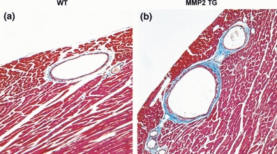
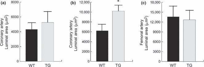
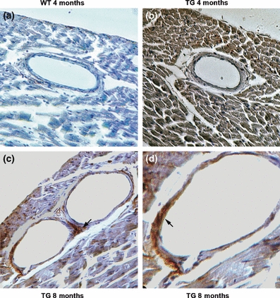
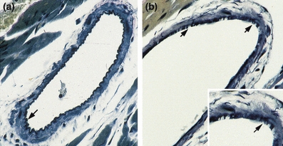
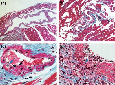
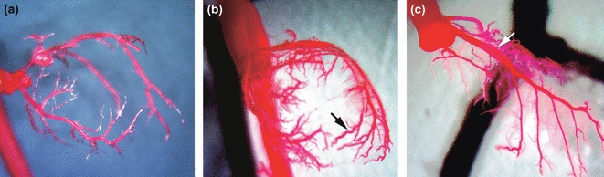
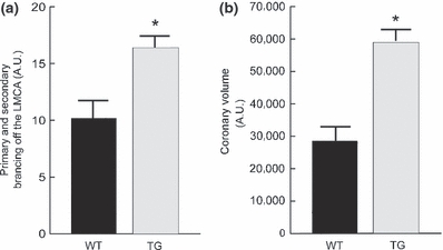
Coronary artery ectasia (CAE) is generally diagnosed in patients undergoing arteriography for presumptive atherosclerotic coronary artery disease. CAE is commonly considered as a variant of atherosclerotic disease; however, recent studies suggest that CAE is the result of a systemic vascular disorder. There is increasing evidence that aneurysmal vascular disease is a systemic disorder characterized by enhanced expression of pro-inflammatory cytokines and increased synthesis of enzymes capable of degrading elastin and other components of the vascular wall. Matrix metalloproteinase-2 degrades a number of extracellular substrates, including elastin and has been shown to play a critical role in the development of abdominal aortic aneurysms. This study characterizes the development of CAE in a unique murine transgenic model with cardiac-specific expression of active MMP-2. Transgenic mice were engineered to express an active form of MMP-2 under control of the α-myosin heavy chain promoter. Coronary artery diameters were quantified, along with studies of arterial structure, elastin integrity and vascular expression of the MMP-2 transgene. Latex casts quantified total coronary artery volumes and arterial branching. Mid-ventricular coronary luminal areas were increased in the MMP-2 transgenics, coupled with foci of aneurysmal dilation, ectasia and perivascular fibrosis. There was no evidence for atherogenesis. Coronary vascular elastin integrity was compromised and coupled with inflammatory cell infiltration. Latex casts of the coronary arteries displayed ectasia with fusiform dilatation. The MMP-2 transgenic closely replicates human CAE and supports a critical and initiating role for this enzyme in the pathogenesis of this disorder.
© 2010 The Authors. International Journal of Experimental Pathology © 2010 International Journal of Experimental Pathology.
Figures
Similar articles
-
Matrix metalloproteinases and inflammatory markers in coronary artery ectasia: their relationship to severity of coronary artery ectasia.Coron Artery Dis. 2008 Dec;19(8):559-63. doi: 10.1097/MCA.0b013e3283109079. Coron Artery Dis. 2008. PMID: 19005290
-
Systemic arterial expression of matrix metalloproteinases 2 and 9 in acute Kawasaki disease.Arterioscler Thromb Vasc Biol. 2003 Apr 1;23(4):576-81. doi: 10.1161/01.ATV.0000065385.47152.FD. Epub 2003 Mar 6. Arterioscler Thromb Vasc Biol. 2003. PMID: 12692003
-
Transcriptional expression profiles of the main proteinases and their regulators in coronary artery ectasia patients' mononuclear cells.Acta Cardiol. 2016 Apr;71(2):157-63. doi: 10.2143/AC.71.2.3141845. Acta Cardiol. 2016. PMID: 27090037
-
Novel insights into an old controversy: is coronary artery ectasia a variant of coronary atherosclerosis?Clin Res Cardiol. 2007 Jun;96(6):331-9. doi: 10.1007/s00392-007-0521-0. Epub 2007 Apr 26. Clin Res Cardiol. 2007. PMID: 17453130 Free PMC article. Review.
-
Pathogenetic mechanisms of coronary ectasia.Int J Cardiol. 2008 Nov 28;130(3):335-43. doi: 10.1016/j.ijcard.2008.05.071. Epub 2008 Aug 9. Int J Cardiol. 2008. PMID: 18694609 Review.
Cited by
-
Plasma Big Endothelin-1 Level Predicted 5-Year Major Adverse Cardiovascular Events in Patients With Coronary Artery Ectasia.Front Cardiovasc Med. 2021 Nov 29;8:768431. doi: 10.3389/fcvm.2021.768431. eCollection 2021. Front Cardiovasc Med. 2021. PMID: 34912865 Free PMC article.
-
A novel intracellular isoform of matrix metalloproteinase-2 induced by oxidative stress activates innate immunity.PLoS One. 2012;7(4):e34177. doi: 10.1371/journal.pone.0034177. Epub 2012 Apr 3. PLoS One. 2012. PMID: 22509276 Free PMC article.
-
Among Ectasia Patients with Coexisting Coronary Artery Disease, TIMI Frame Count Correlates with Ectasia Size and Markis Type IV Is the Commonest.Cardiol Res Pract. 2015;2015:282170. doi: 10.1155/2015/282170. Epub 2015 Feb 3. Cardiol Res Pract. 2015. PMID: 25705544 Free PMC article.
-
Prevalence of coronary artery ectasia in older adults and the relationship with epicardial fat volume by cardiac computed tomography angiography.J Geriatr Cardiol. 2013 Mar;10(1):10-5. doi: 10.3969/j.issn.1671-5411.2013.01.003. J Geriatr Cardiol. 2013. PMID: 23610568 Free PMC article.
-
The Impact of C-Peptide and Diabetes Mellitus on Coronary Ectasia and Effect of Coronary Ectasia and C-Peptide on Long-Term Outcomes: A Retrospective Cohort Study.Int J Clin Pract. 2022 Oct 8;2022:7910566. doi: 10.1155/2022/7910566. eCollection 2022. Int J Clin Pract. 2022. PMID: 36277470 Free PMC article.
References
-
- Alfonso-Jaume MA, Bergman MR, Mahimkar R, et al. Cardiac ischemia-reperfusion injury induces matrix metalloproteinase-2 expression through the AP-1 components FosB and JunB. Am. J. Physiol. Heart Circ. Physiol. 2006;291:H1838–H1846. - PubMed
-
- Bergman MR, Teerlink JR, Mahimkar R, et al. Cardiac matrix metalloproteinase-2 expression independently induces marked ventricular remodeling and systolic dysfunction. Am. J. Physiol. Heart Circ. Physiol. 2007;292:H1847–H1860. - PubMed
-
- Cai WJ, Kocsis E, Wu X, et al. Remodeling of the vascular tunica media is essential for development of collateral vessels in the canine heart. Mol. Cell. Biochem. 2004;264:201–210. - PubMed
-
- Davis V, Persidskaia R, Baca-Regen L, et al. Matrix metalloproteinase-2 production and its binding to the matrix are increased in abdominal aortic aneurysms. Arterioscler. Thromb. Vasc. Biol. 1998;18:1625–1633. - PubMed
-
- Finkelstein A, Michowitz Y, Abashidze A, Miller H, Keren G, George J. Temporal association between circulating proteolytic, inflammatory and neurohormonal markers in patients with coronary ectasia. Atherosclerosis. 2005;179:353–359. - PubMed
Publication types
MeSH terms
Substances
Grants and funding
LinkOut - more resources
Full Text Sources
Other Literature Sources
Medical
Miscellaneous
