. 2011 Feb;92(1):26-39.
doi: 10.1111/j.1365-2613.2010.00746.x. Epub 2010 Oct 29.

Combining two potential causes of metalloproteinase secretion causes abdominal aortic aneurysms in rats: a new experimental model

Affiliations

Combining two potential causes of metalloproteinase secretion causes abdominal aortic aneurysms in rats: a new experimental model

Karina M Mata et al. Int J Exp Pathol. 2011 Feb.

Abstract

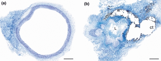
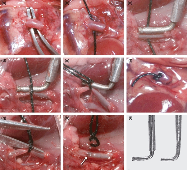
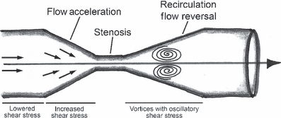
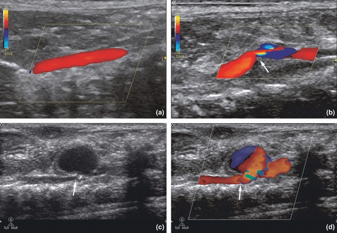
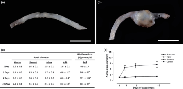
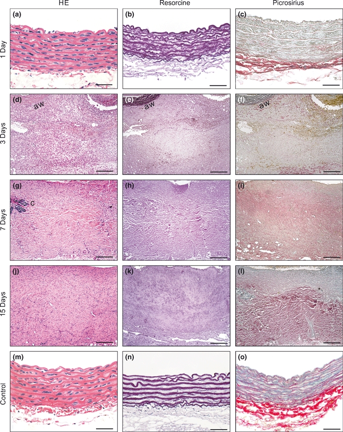
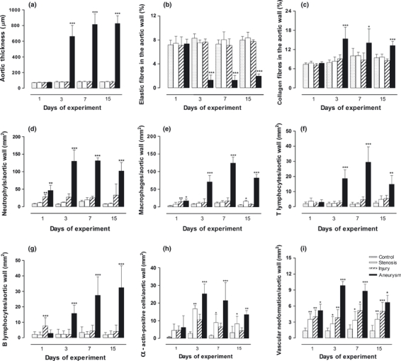
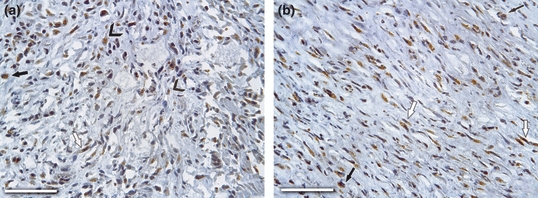
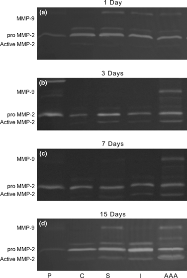
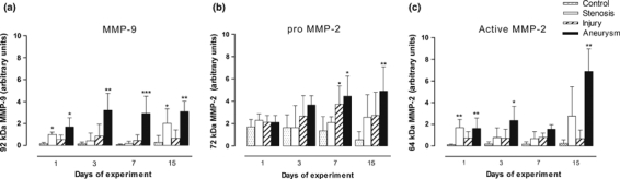
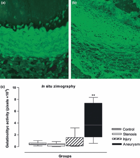
Progress in understanding the pathophysiology of abdominal aortic aneurysms (AAA) is dependent in part on the development and application of effective animal models that recapitulate key aspects of the disease. The objective was to produce an experimental model of AAA in rats by combining two potential causes of metalloproteinase (MMP) secretion: inflammation and turbulent blood flow. Male Wistar rats were randomly divided in four groups: Injury, Stenosis, Aneurysm and Control (40/group). The Injury group received a traumatic injury to the external aortic wall. The Stenosis group received an extrinsic stenosis at a corresponding location. The Aneurysm group received both the injury and stenosis simultaneously, and the Control group received a sham operation. Animals were euthanized at days 1, 3, 7 and 15. Aorta and/or aneurysms were collected and the fragments were fixed for morphologic, immunohistochemistry and morphometric analyses or frozen for MMP assays. AAAs had developed by day 3 in 60-70% of the animals, reaching an aortic dilatation ratio of more than 300%, exhibiting intense wall remodelling initiated at the adventitia and characterized by an obvious inflammatory infiltrate, mesenchymal proliferation, neoangiogenesis, elastin degradation and collagen deposition. Immunohistochemistry and zymography studies displayed significantly increased expressions of MMP-2 and MMP-9 in aneurysm walls compared to other groups. The haemo-dynamic alterations caused by the stenosis may have provided additional contribution to the MMPs liberation. This new model illustrated that AAA can be multifactorial and confirmed the key roles of MMP-2 and MMP-9 in this dynamic remodelling process.


Figures

Figure 1
Figure 1
Prestenotic luminal aorta (a) compared with the most stenotic luminal area (b). Note the extreme reduction of lumen (L) because of the ligature of the aorta with cotton thread (ct) (HRLM, Toluidine blue). Bar = 200 μm.
Figure 2
Figure 2
Photographic sequence of the surgical procedure used in the experiment. (a) Control group: only mobilization of the aorta was carried out; (a–f) Aneurysm group: after dissection, a dental diamond bur is placed over the aorta (c). A ligature of cotton thread is made around the bur (d), which is immediately removed (e), reducing the vessel lumen to the diameter of the probe (f); (a–h) Injury group: the ligature was cut after the bur diamond was withdrawn (g, h), provoking wall injury (arrow), and (i): At left, a stainless steel version of a dental bur diamond and at right the tool without the bur diamond.
Figure 3
Figure 3
Schematic representation of the shear stress patterns induced by stenosis is show (Modified from Cheng et al. 2006).
Figure 4
Figure 4
Colour Doppler: (a) Dark red near the aorta wall, representing laminar flow in the abdominal aortas from Injury and Control groups. (b) Stenotic group showing laminar flow, which appears dark-orange to yellow in the prestenotic segment and mixed colours in the poststenotic segment, indicating turbulent flow. The arrow points the stenosis in the aorta. Grey scale (c) and colour Doppler (d) of the (a) group on the third day. Note the abdominal aorta with a narrowing (arrows) is associated with a true aneurysm. The mixed colour within the aneurysm represents turbulent flow.
Figure 5
Figure 5
Macroscopic view of the abdominal aortas: (a) Representative aortas from the Stenosis and Injury groups showing no abnormal dilatations, and (b) a large aneurysm with a dilatation ratio of more than 300% restricted to the previously injured-stenosed area observed on the third day in the Aneurysm group. Bar = 1 cm. (c) Table showing the mean aortic diameters of the groups (mm) and the dilatation ratio in the Aneurysm group during the experiment. (d) Change in aortic diameter during the experiment. The data are shown as the means ± SD (n=6 per group; a: P < 0.001).
Figure 6
Figure 6
Microscopic views of the Aneurysm group at each experimental time point (a–l). On the first day, no significant alterations were observed. On the other days, extensive wall remodelling moving into the adventitial tissue was seen, characterized by degeneration of extracellular matrix, mesenchymal cell proliferation, increased angiogenesis and abundant collagen deposition progressing inward towards the whole aortic wall (aw). Note the massive elastic fibre destruction and fragmentation, prominent starting on the third day. Calcification foci (c) were seen. At 7 and 15 dps, the alterations were similar to those at 3 dps, except for the greater wall thickness, less inflammatory infiltration and neovascular formation, and apparently denser collagen deposition. Representative microscopic examples of the aortas of the Control, Stenosis and Injury groups without significant alterations are shown (m–o).Bar = 25 μm (a–c, m–o), 135 μm (d–l). Original magnification, 400×.
Figure 7
Figure 7
Bar graphs showing the changes in the different morphometric parameters analysed in all groups. Data shown are means ± SD (n=6 per group); *P < 0.05, **P < 0.01 and ***P < 0.001 [Aneurysm group vs. Control, Stenosis and Injury groups in a–c, f and g; Stenosis group vs. Control and Injury groups in e, h and i, and Injury group vs. Control and Stenosis groups in d, e and i].
Figure 8
Figure 8
Immunohistochemical localization of MMP-9 and MMP-2 in aneurismal wall on day 3 after surgery are show. (a) MMP-9 showed strong expression throughout the remodelling wall, especially within neutrophils (arrowheads), macrophages (arrow) and myofibroblasts (open arrow), and (b) MMP-2 were strongly marked in macrophages (arrows) and myofibroblasts (open arrows). Bar = 150 μm; Original magnification, 400×.
Figure 9
Figure 9
Representative SDS–PAGE gelatin zymography of aortic samples (a–d), showing increased expression of MMP-9, identified as a 92 kDa band, and latent (72 kDa) and active forms (64 kDa) of MMP-2 in the aneurysms extracts (AAA) in all days after surgery. P, pattern; C, Control; S, Stenosis; I, Injury; AAA, Aneurysm groups.
Figure 10
Figure 10
Bar graphs show the densitometric analyses of the MMP-2 and MMP-9 activities during the experiment. (a) Note the elevated levels of the MMP-9 in the Aneurysm group, starting at the first dps and remaining elevated at similar levels until the end of the experiment. (b) Pro-MMP-2 was significantly expressed after the seventh dps, especially in the aneurysms groups. (c) Active MMP-2 increased expression showed a similar behaviour to the latent form in the aneurysm group, with significant levels on most days of the experiment. *P < 0.05, **P < 0.01 and ***P < 0.001 [Aneurysm group vs. Injury, Stenosis and Control groups (a–c); Stenosis group vs. Injury and Control groups (a, c) and, Injury group vs. Stenosis and Control groups (b)].
Figure 11
Figure 11
Representative images of in situ zymography of the aortic wall as seen in the control group (a) and in the aneurysmal group (b) at 3 dps. Note the intense fluorescence (light green) indicating gelatinolytic activity and wall degradation (black areas) in the aneurysmal wall (b). Original magnification, 400×. (c) Boxplot of optical density by image analysis showing the intense gelatinolytic activity in the aneurysmal wall at the third day of the experiment. **P < 0.01 [Anerysm group vs. Control, Stenosis and Injury groups].

Similar articles

Cited by

References

    1. Allaire E, Forough R, Clowes M, Starchcr B, Clowes AW. Local over expression of TIMP-1 prevents aortic aneurysm degeneration and rupture in a rat model. J. Clin. Invest. 1998;102:1413–1420. - PMC - PubMed
    1. Anidjar S, Salzmann JL, Gentric D, Lagneau P, Camilleri JP, Michel JB. Elastase-induced experimental aneurysms in rats. Circulation. 1990;82:973–981. - PubMed
    1. Aoki H, Yoshimura K, Matsuzaki M. Turning back the clock: regression of abdominal aortic aneurysms via pharmacotherapy. J. Mol. Med. 2007;85:1077–1088. - PubMed
    1. Basalyga DM, Simionescu DT, Xiong W, Baxter BT, Starcher BC, Vyavahare NR. Elastin degradation and calcification in an abdominal aorta injury model: role of matrix metalloproteinases. Circulation. 2004;110:3480–3487. - PMC - PubMed
    1. Bigatel DA, Elmorc JR, Carey DJ, Cizlncci SG, Franklin DR, Youkcy JR. The matrix metalloproteinase inhibitor BB-94 lintits expansion of experimental abdontinal aortic ancuwsms. J. Vasc. Surg. 1999;29:130–138. - PubMed

Publication types

Substances

LinkOut - more resources