

Oleic acid ameliorates amyloidosis in cellular and mouse models of Alzheimer's disease
- PMID: 21040071
- PMCID: PMC8094298
- DOI: 10.1111/j.1750-3639.2010.00449.x
Oleic acid ameliorates amyloidosis in cellular and mouse models of Alzheimer's disease
Erratum in
-
Erratum.Brain Pathol. 2017 Sep;27(5):702-703. doi: 10.1111/bpa.12512. Brain Pathol. 2017. PMID: 28805008 Free PMC article. No abstract available.
Abstract
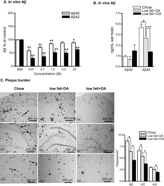
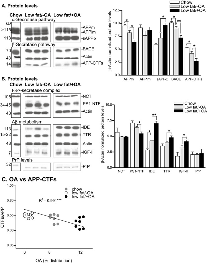
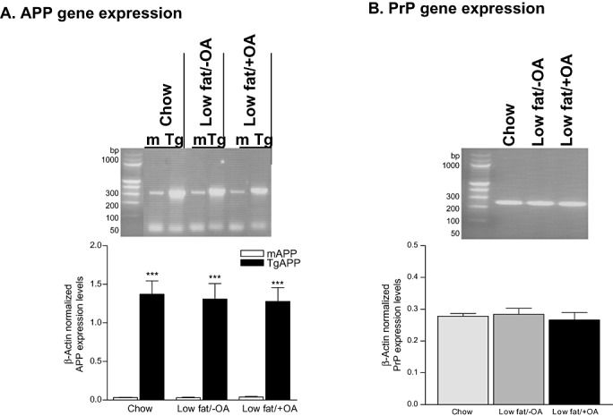
Several lines of evidence support protective as well as deleterious effects of oleic acid (OA) on Alzheimer's disease (AD) and other neurological disorders; however, the bases of these effects are unclear. Our investigation demonstrates that amyloid precursor protein (APP) 695 transfected Cos-7 cells supplemented with OA have reduced secreted amyloid-beta (Aβ) levels. An early-onset AD transgenic mouse model expressing the double-mutant form of human APP, Swedish (K670N/M671L) and Indiana (V717F), corroborated our in vitro findings when they were fed a high-protein, low-fat (18% reduction), cholesterol-free diet enriched with OA. These mice exhibited an increase in Aβ40/Aβ42 ratio, reduced levels of beta-site APP cleaving enzyme (BACE) and reduced presenilin levels along with reduced amyloid plaques in the brain. The decrease in BACE levels was accompanied by increased levels of a non-amyloidogenic soluble form of APP (sAPPα). Furthermore, the low-fat/+OA diet resulted in an augmentation of insulin-degrading enzyme and insulin-like growth factor-II. These results suggest that OA supplementation and cholesterol intake restriction in a mouse model of AD reduce AD-type neuropathology.
© 2010 The Authors; Brain Pathology © 2010 International Society of Neuropathology.
Figures
References
-
- Ahren B (1999) Plasma leptin and insulin in C57BI/6J mice on a high‐fat diet: relation to subsequent changes in body weight. Acta Physiol Scand 165:233–240. - PubMed
-
- Amtul Z, Uhrig M, Supino R, Beyreuther K (2010) Phospholipids and a phospholipid‐rich diet alter the in vitro amyloid‐beta peptide levels and amyloid‐beta 42/40 ratios. Neurosci Lett 481:73–77. - PubMed
-
- Aoyagi T, Wada T, Nagai M, Kojima F, Harada S, Takeuchi T et al (1990) Deficiency of kallikrein‐like enzyme activities in cerebral tissue of patients with Alzheimer's disease. Experientia 46:94–97. - PubMed
-
- Askari B, Carroll MA, Capparelli M, Kramer F, Gerrity RG, Bornfeldt KE (2002) Oleate and linoleate enhance the growth‐promoting effects of insulin‐like growth factor‐I through a phospholipase d‐dependent pathway in arterial smooth muscle cells. J Biol Chem 277:36338–36344. - PubMed
-
- Brijbassi S, Amtul Z, Newbigging S, Westaway D, St George‐Hyslop P, Rozmahel RF (2007) Excess of nicastrin in brain results in heterozygosity having no effect on endogenous APP processing and amyloid peptide levels in vivo . Neurobiol Dis 25:291–296. - PubMed
Publication types
MeSH terms
Substances
Grants and funding
LinkOut - more resources
Full Text Sources
Medical
