. 2010 Oct;17(7):536-47.
doi: 10.1111/j.1549-8719.2010.00047.x.

Murine rVEGF164b, an inhibitory VEGF reduces VEGF-A-dependent endothelial proliferation and barrier dysfunction

Affiliations

Murine rVEGF164b, an inhibitory VEGF reduces VEGF-A-dependent endothelial proliferation and barrier dysfunction

Walter Cromer et al. Microcirculation. 2010 Oct.

Erratum in

  • Microcirculation. 2010 Nov;17(8):669

Abstract

Objective: To investigate the effects of the murine inhibitory vascular endothelial growth factor (VEGF, rVEGF164b), we generated an adenoviral vector encoding rVEGF164b, and examined its effects on endothelial barrier, growth, and structure.

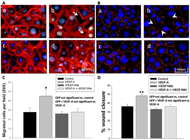
Method: Mouse vascular endothelial cells (MVEC) proliferation was determined by an MTT assay. Barrier of MVEC monolayers was measured by trans-endothelial electrical resistance (TEER). Reorganization of actin and zonula occludens-1 (ZO-1) were determined by fluorescent microscopy.

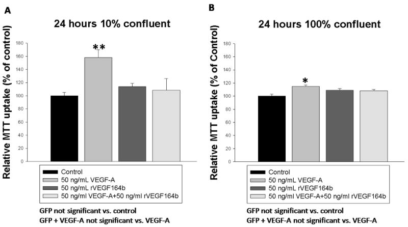
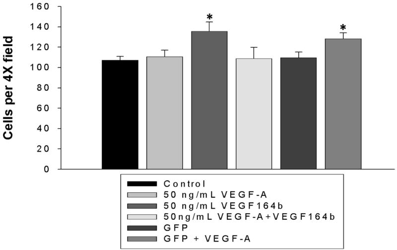
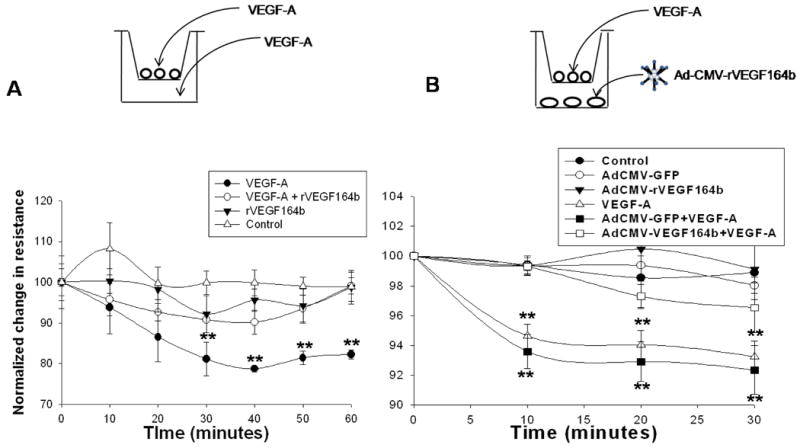
Results: Mouse venous endothelial cells treated with murine VEGF-A (VEGF-A) (50 ng/mL) increased proliferation (60.7 ± 0.1%) within 24 hours (p < 0.05) and rVEGF164b inhibited VEGF-A-induced proliferation. TEER was significantly decreased by VEGF-A (81.7 ± 6.2% of control). Treatment with rVEGF164b at 50 ng/mL transiently reduced MVEC barrier (p < 0.05) at 30 minutes post-treatment (87.9 ± 1.7% of control TEER), and returned to control levels by 40 minutes post-treatment. Treatment with rVEGF164b prevented barrier changes by subsequent exposure to VEGF-A. Treatment of MVECS with VEGF-A reorganized F-actin and ZO-1, which was attenuated by rVEGF164b.

Conclusions: VEGF-A may dysregulate endothelial barrier through junctional cytoskeleton processes, which can be attenuated by rVEGF164b. The VEGF-A stimulated MVEC proliferation, barrier dysregulation, and cytoskeletal rearrangement. However, rVEGF164b blocks these effects, therefore it may be useful for regulation studies of VEGF-A/VEGF-R signaling in many different models.


Figures

Figure 1
Figure 1
(A) Sequence alignment of the predicted COOH terminal of VEGF164b with rVEGF164b, VEGF165b, and VEGF165. (B) Grantham polarity plots of the COOH terminal end of VEGF-A, VEGF165b, and rVEGF164b. (C) Hydrophobicity plots of the COOH terminal of VEGF-A, VEGF165b, and rVEGF164b.
Figure 2
Figure 2
(A) Denaturing Western blot of rVEGF164b from A549 cells 48 h post-infection showing 50ng of native murine VEGF-A protein (lane 1) and 50ng rVEGF164b in conditioned medium (lane 2). Both bands appear at 21kDa as expected, the higher molecular weight band is non-specific interaction of the antibody with BSA. (B) Non-denaturing Western blot of Ad5-CMV-rVEGF164b infected YAMCs (lane 1) and uninfected cells (lane 2). Bands appear at 42kDa which corresponds to a VEGF dimer pair (biologically active form) of the protein. (C) Production of rVEGF164b in A549 measured by ELISA. Total VEGF accumulation in media represents total accumulated VEGF in media at the corresponding time point and VEGF production per 24 hour period shows total VEGF production during the indicated 24 hour time point. Bars are means ± standard error of the mean (SEM) of three independent measurements.
Figure 3
Figure 3
(A) Inhibition of VEGF-A induced proliferation by rVEGF164b in MVECs seeded at 10% confluence at 24 hours as measured by MTT conversion (n = 32). (B) Measurement of VEGF-A induced mitochondrial activity by MTT conversion in fully confluent MVECs and inhibition by rVEGF164b (n=16). Bars are means ± SEM of three independent measurements. Results were analyzed by one-way ANOVA with Dunnett's post-hoc test. *p < 0.05.
Figure 4
Figure 4
Cells seeded at low density (100 cells/0.33cm2) were treated with media containing VEGF-A, rVEGF164b, a combination of the two or control media. Cell number was counted at low power (4×). rVEGF164b significantly inhibited VEGF-A induced proliferation. Bars are means ± SEM of three independent measurements. Results were analyzed by one-way ANOVA with Dunnett's post-hoc test. *p < 0.05.
Figure 5
Figure 5
(A) Trans-Endothelial Electrical Resistance (TEER) of MVECs testing for inhibition of VEGF-A mediated decrease in barrier function. Resistance was significantly decreased by VEGF-A for 60 min post treatment and did not return to normal for at least 120 min. Resistance returned to normal in rVEGF164b and VEGF-A + rVEGF164b treated monolayers at 60 min post-treatment. Bars are means ± SEM of three independent measurements. Results were analyzed by repeated measures ANOVA with Dunnett's post-hoc test for deviation from resistance at time = 0. *p < 0.05, n = 3. (B) TEER readings of MVECs cultured above YAMCs infected with 100:1 titer of Ad5-CMV-rVEGF164b following a 50 ng/mL spike of VEGF-A at 48 h. Bars are means ± SEM of three independent measurements. Protection was determined by repeated measures ANOVA with Dunnett's post-hoc test. *p < 0.05, **p < 0.01, n = 6.
Figure 6
Figure 6
(A) Rhodamine phalloidin staining of confluent MVECs after 45 min. treatment with (a) vehicle, (b) VEGF-A, (c) rVEGF164b, or (d) co-treatment with rVEGF164b and VEGF-A. Apparent gaps are marked by arrows (white). Each image is representative of three separate experiments. (B) ZO-1 immunofluorescence staining of confluent MVECs after 2 h treatment with (a) vehicle, (b) VEGF-A, (c) rVEGF164b, or (d) co-treatment with rVEGF164b and VEGF-A. Each image is representative of four separate experiments. (C) Transwell migration assay of MVECs in which VEGF-A induced significant migration across insert membranes and rVEGF164b inhibited this chemotactic effect while no protective effect was seen in GFP contols n=3. Bars are means ± SEM of three independent measurements. Significance was determined by ANOVA with Dunnett's post-hoc test. *p < 0.05. (D) MVECs were allowed to grow to confluence in 96 well dishes at which time growth medium was switched to starvation medium for 48 hours. Cell monolayers were wounded with the point of a 1000μL pipette tip and the starvation medium was removed and replaced with experimental media and allowed to migrate for 18 h. Percent wound closure was calculated and statistical significance was determined by ANOVA with Dunnett's post test. Bars are ± SEM, n=4. *p<0.01.

References

    1. Woolard J, Wang WY, Bevan HS, Qiu Y, Morbidelli L, Pritchard-Jones RO, Cui TG, Sugiono M, Waine E, Perrin R, et al. VEGF165b, an inhibitory vascular endothelial growth factor splice variant: mechanism of action, in vivo effect on angiogenesis and endogenous protein expression. Cancer Res. 2004;64(21):7822–35. - PubMed
    1. Olsson AK, Dimberg A, Kreuger J, Claesson-Welsh L. VEGF receptor signalling - in control of vascular function. Nat Rev Mol Cell Biol. 2006;7(5):359–71. - PubMed
    1. Appleton I, Brown NJ, Willis D, Colville-Nash PR, Alam C, Brown JR, Willoughby DA. The role of vascular endothelial growth factor in a murine chronic granulomatous tissue air pouch model of angiogenesis. J Pathol. 1996;180(1):90–4. - PubMed
    1. Ten Brick Robin A, C DT, Martin Jeremy L, Moline Karl V, Bergman Jeffery W, Cupp Andrea S. Vascular Endothelial Growth Factor Inhibitory Isoform Is Regulated Prior to Ovulation. Nebraska Beef Cattle Reports. 2007;1(1):113–115.
    1. Usui T, Ishida S, Yamashiro K, Kaji Y, Poulaki V, Moore J, Moore T, Amano S, Horikawa Y, Dartt D, et al. VEGF164(165) as the pathological isoform: differential leukocyte and endothelial responses through VEGFR1 and VEGFR2. Invest Ophthalmol Vis Sci. 2004;45(2):368–74. - PubMed

Publication types

MeSH terms