

Unraveling the Golgi ribbon
- PMID: 21040294
- PMCID: PMC4221251
- DOI: 10.1111/j.1600-0854.2010.01114.x
Unraveling the Golgi ribbon
Abstract
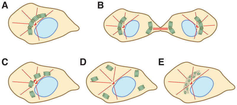
The Golgi apparatus lies at the heart of the secretory pathway where it receives, modifies and sorts protein cargo to the proper intracellular or extracellular location. Although this secretory function is highly conserved throughout the eukaryotic kingdom, the structure of the Golgi complex is arranged very differently among species. In particular, Golgi membranes in vertebrate cells are integrated into a single compact entity termed the Golgi ribbon that is normally localized in the perinuclear area and in close vicinity to the centrosomes. This organization poses a challenge for cell division when the single Golgi ribbon needs to be partitioned into the two daughter cells. To ensure faithful inheritance in the progeny, the Golgi ribbon is divided in three consecutive steps in mitosis, namely disassembly, partitioning and reassembly. However, the structure of the Golgi ribbon is only present in higher animals and Golgi disassembly during mitosis is not ubiquitous in all organisms. Therefore, there must be unique reasons to build up the Golgi in this particular conformation and to preserve it over generations. In this review, we first highlight the diversity of the Golgi architecture in different organisms and revisit the concept of the Golgi ribbon. Following on, we discuss why the ribbon is needed and how it forms in vertebrate cells. Lastly, we conclude with likely purposes of mitotic ribbon disassembly and further propose mechanisms by which it regulates mitosis.
© 2010 John Wiley & Sons A/S.
Figures
References
-
- Mowbrey K, Dacks JB. Evolution and diversity of the Golgi body. FEBS Lett. 2009;583:3738–3745. - PubMed
-
- Rambourg A, Jackson CL, Clermont Y. Three dimensional configuration of the secretory pathway and segregation of secretion granules in the yeast Saccharomyces cerevisiae. J Cell Sci. 2001;114:2231–2239. - PubMed
-
- Puthenveedu MA, Bachert C, Puri S, Lanni F, Linstedt AD. GM130 and GRASP65-dependent lateral cisternal fusion allows uniform Golgi-enzyme distribution. Nat Cell Biol. 2006;8:238–248. - PubMed
Publication types
MeSH terms
Grants and funding
LinkOut - more resources
Full Text Sources
