. 2010 Nov-Dec;12(6):552-67.
doi: 10.1111/j.1525-142X.2010.00441.x.

Phylogeny informs ontogeny: a proposed common theme in the arterial pole of the vertebrate heart

Affiliations

Phylogeny informs ontogeny: a proposed common theme in the arterial pole of the vertebrate heart

Adrian C Grimes et al. Evol Dev. 2010 Nov-Dec.

Abstract

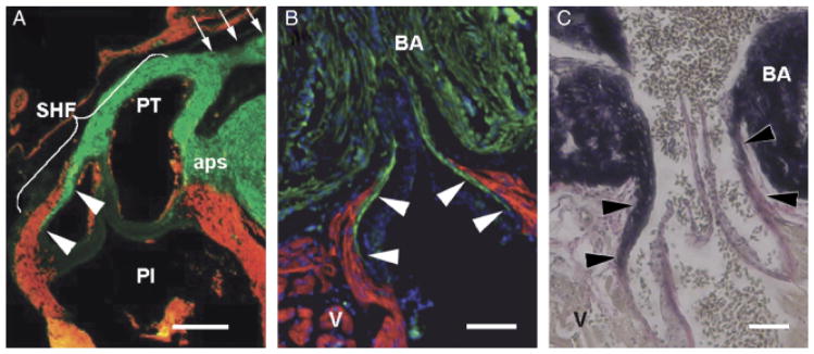
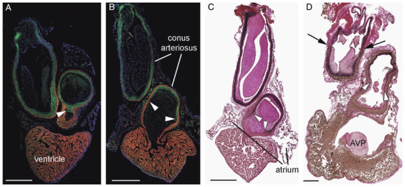
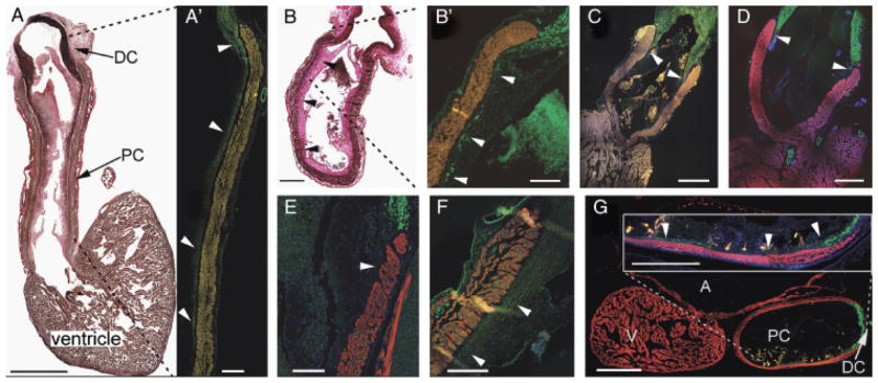
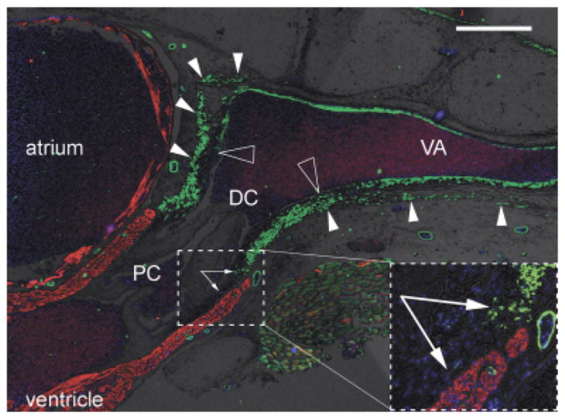
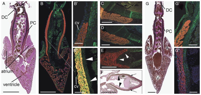
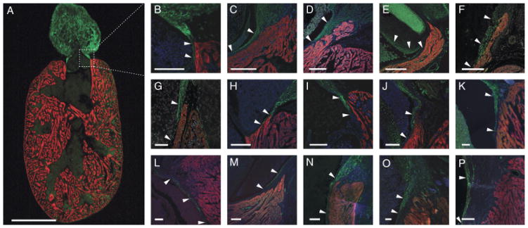
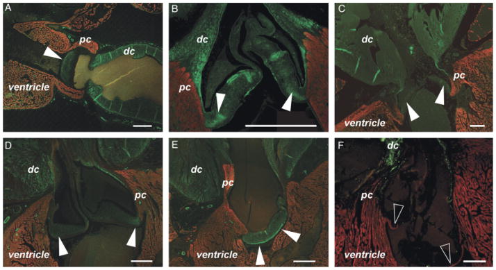
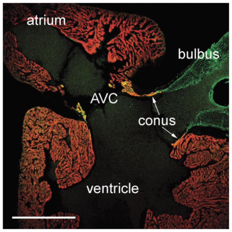
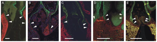
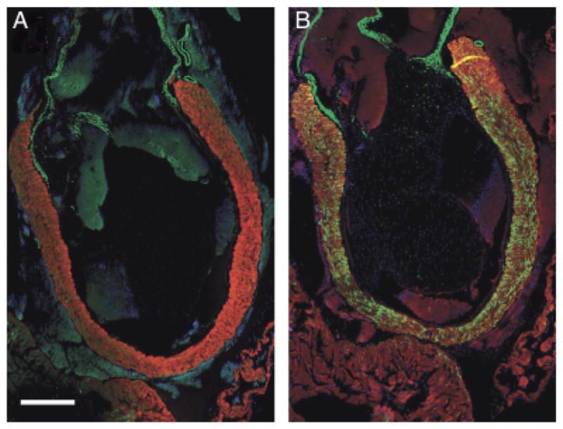
In chick and mouse embryogenesis, a population of cells described as the secondary heart field (SHF) adds both myocardium and smooth muscle to the developing cardiac outflow tract (OFT). Following this addition, at approximately HH stage 22 in chick embryos, for example, the SHF can be identified architecturally by an overlapping seam at the arterial pole, where beating myocardium forms a junction with the smooth muscle of the arterial system. Previously, using either immunohistochemistry or nitric oxide indicators such as diaminofluorescein 2-diacetate, we have shown that a similar overlapping architecture also exists in the arterial pole of zebrafish and some shark species. However, although recent work suggests that development of the zebrafish OFT may also proceed by addition of a SHF-like population of cells, the presence of a true SHF in zebrafish and in many other developmental biological models remains an open question. We performed a comprehensive morphological study of the OFT of a wide range of vertebrates. Our data suggest that all vertebrates possess three fundamental OFT components: a proximal myocardial component, a distal smooth muscle component, and a middle component that contains overlapping myocardium and smooth muscle surrounding and supporting the outflow valves. Because the middle OFT component of avians and mammals is derived from the SHF, our observations suggest that a SHF may be an evolutionarily conserved theme in vertebrate embryogenesis.


Figures

Fig. 1
Fig. 1
Architectural homology in the arterial pole of two widely diverged species. In all three images, cranial is to the top. (A) Detail of the chick pulmonary outflow, gestational day 11, sectioned longitudinally and labeled with MF20 (myocardium—red) and anti-SMA (smooth muscle—green). The pulmonary outflow valves are surrounded and supported by a collar of myocardium. Smooth muscle known to be derived from the secondary heart field (SHF) can be seen at the lumenal face of the myocardial collar, extending caudally from the base of the arterial trunk to the level of the valves (arrowheads). Arrows show distal outflow tract smooth muscle known to be neural crest derived. The smooth muscle in the remnant of the aorticopulmonary septum (aps) is also neural crest derived. PI, pulmonary infundibulum; PT, pulmonary trunk. Scale bar=250 μm (image adapted from Waldo et al. 2005). (B) Detail of the adult zebrafish arterial pole sectioned longitudinally and labeled with MF-20 (red) and anti-MLCK (smooth muscle—green). DAPI (blue) labels nuclei. White arrowheads show that, as in the chick, the valves are supported by a collar of myocardium (the conus arteriosus) and that smooth muscle extends caudally from the base of the bulbus arteriosus (BA) to the level of the valve leaflets. Scale bar=100 μm. (C) Similar longitudinal section of adult zebrafish stained with elastic trichrome, showing an abundance of fibrous and elastic proteins (arrowheads) in the “fibrous ring” connecting the ventricle (V) with the bulbus arteriosus (BA). Scale bar=100 μm.
Fig. 2
Fig. 2
Confirmation of a smooth muscle-myocardium overlap in lungfish species. African (slender) lungfish Protopterus dolloi (A) and South American lungish Lepidosiren paradoxa (B) labeled with CH1 (myocardium—red) and SM22α (green). Cell nuclei are labeled with DAPI (blue). In each image, cranial is to the top. The smooth muscle of the outflow tract (OFT) extends to the base of the endocardial valves (arrowheads). The myocardial collar extends almost as far as the pericardial wall. Note however that a small portion of the distal OFT contains no myocardium. (C and D) Elastic trichrome stain of Lepidosiren paradoxa (B) and the Australian lungfish Neoceratodus forsteri. Note that similar to in the zebrafish, elastic and fibrous proteins (stained black) coincide with the smooth muscle cells almost to the proximal limit of the valves (arrowheads). Black arrows in (D) show the distal intrapericardial limit of the conal myocardium. AVP, atrioventricular plug. Scale bars=1 mm.
Fig. 3
Fig. 3
Chondrichthyans exhibit similar arterial pole architechture. (A) Sagittal section through the heart of Dalatias licha. Scale bar=5 mm. Cranial to the top, dorsal to the left—elastic trichrome stains muscle brown, collagen red, and elastic proteins black. Abundant elastic fibers in the distal smooth muscle component of the outflow tract (OFT) extend caudally into the subendocardium as far as the most proximal valve leaflets. (A′) Detail of the ventral portion of the OFT marked by the dashed lines, labeled with anti-SM22α (green). Scale bar=1 mm. Arrowheads show that smooth muscle is coincident with elastic fibers in the subendocardial layer. Autofluorescence in the myocardium has been artificially enhanced. (B) Longitudinal section through the OFT of Etmopterus spinax—elastic trichrome. Scale bar=500 μm. Subendocardial elastic fibers (arrowheads) extend to the level of the most proximal valve leaflets. (B′) Detail of the region marked by dashed lines in (B). Anti-SMA shows coincident subendocardial smooth muscle (arrowheads) in E. spinax. Scale bar=250 μm. Smooth muscle (arrowheads) extends to the most proximal valve. (C and D) The OFT of two Galeus spp., G. atlanticus, and G. melastomus, labeled with CH1 (red) and SM22α (green). Scale bars=1 mm. Smooth muscle (arrowheads) extends to the most distal valves. (E) Detail of the arterial pole of Scyliorhinus canicula. Scale bar=500 μm. The dorsal wall of the conus is shown in sagittal section. CH1 (red) and anti-SMA (green). In this species, the overlapping region is not so extensive, barely reaching the most distal valves (arrowheads). (F) Smooth muscle (arrowheads) extends beyond the distal valves in Leucoraja naevus. CH1 (red), SM22α (green). Scale bar=250 μm. (G) Sagittal section of the heart of Chimaera monstrosa—CH1 (red), SM22α (green). Scale bar=500 μm. Inset shows detail of the ventral OFT wall and that smooth muscle extends to the most proximal valves (arrowheads). Scale bar=250 μm. A, atrium; DC, distal smooth muscle component (bulbus arteriosus); PC, proximal myocardial component (conus arteriosus); V, ventricle.
Fig. 4
Fig. 4
Distinct origin of smooth muscle in distal OFT and ventral aorta? Sagittal section through the heart, outflow tract (OFT) and ventral aorta of a juvenile small-spotted catshark Scyliorhinus canicula, labeled with anti-SMA (green), CH1 (red), and DAPI (blue). Brightfield background shows gross tissue structure. Dorsal to the top, cranial to the right. Large hollow arrowheads show the distal limit of the intrapericardial OFT smooth muscle and, more cranially, the termination of most of the smooth muscle in the tunica media of the ventral aorta. Small arrowheads show where the OFT smooth muscle is continuous with smooth muscle in the pericardium/body wall (most obvious dorsally). Arrows in inset show smooth muscle cells in the subendocardium of the conus arteriosus. Scale bar=500 μm. DC, distal smooth muscle component (bulbus arteriosus); PC, proximal myocardial component (conus arteriosus); VA, ventral aorta.
Fig. 5
Fig. 5
Basal actinopterygians. In all panels showing immunofluorescence, myocardium is labeled with CH1 (red) and, unless otherwise stated, smooth muscle is labeled with anti-SM22α (green). (A) Longitudinal section through the heart of the bichir Polypterus senegalus. Elastic stain shows abundant elastic fibers extending from the distal smooth muscle component (DC) of the outflow tract (OFT) deep into the subendocardium of the proximal component (PC). Scale bar=1 mm. (B) Consecutive section of P. senegalus, anti-SMA (green) and DAPI (blue). Scale bar=1 mm. (B′) Detail of the transition between proximal and distal OFT components showing myocardium-smooth muscle overlap. Scale bar=100 μm. CV, coronary vessel, also labeled with anti-SMA. (B″) Detail of one lateral wall of the proximal OFT component of P. senegalus. Scale bar=100 μm. Dashed line from (B) illustrates approximate relative position of the two sections. Note that the myocardium is also labeled with anti-SM22α, resulting in yellow fluorescence (see “Discussion”). White arrowheads show abundant smooth muscle cells in the subendocardial space. (C and D) Detail of the transition between proximal and distal OFT components of the sturgeons Acipenser stellatus and Acipenser baeri, respectively. Scale bars=500 μm. (E) The OFT of the American paddlefish Polyodon spathula. Scale bar=200 μm. Anti-SMA (green) highlights smooth muscle in the subendocardium (arrowheads). (F) The OFT of the bowfin Amia calva. Scale bar=1 mm. Elastic stain shows elastic fibers deep within the subendocardium (arrowheads). Black arrows show the distal limit of the myocardial OFT component. (G) The heart of Lepisosteus osseus—elastic staining. Scale bar=500 μm. A, atrium; DC, distal smooth muscle component (bulbus arteriosus); PC, proximal myocardial component (conus arteriosus); V, ventricle. (G′) Detail of the transition between proximal and distal OFT components of L. osseus. Scale bar=150 μm. (G″) Detail of L. osseus proximal component wall. Dashed line from (G) shows approximate relative position of the two sections. Scale bar=150 μm.
Fig. 6
Fig. 6
Myocardial—smooth muscle overlap in the teleost arterial pole. (A) Longitudinal section through the heart of Xiphophorus helleri, a representative teleost, labeled with CH1 (red) and anti-MLCK (green). Cranial to the top. Scale bar=500 μm. (B) Detail of one side of the arterial pole (boxed section in A) showing smooth muscle extending caudally into the myocardial region supporting the outflow valves (the conus arteriosus). Scale bar=100 μm. (C–P) Details of similar regions in the arterial pole of 14 representative teleost species. Scale bars=100 μm. Giant snakehead Channa micropeltes (C), walleye Sander vitreus (D), silver arowana Osteoglossum bicirrhosum (E), black ghost knife fish Apteronotus albifrons (F), gar characin Ctenolucius hujeta (G), Altlantic silversideMenidia menidia (H), suckermouth catfish Hypostomus plecostomus (I), European pilchard Sardina pilchardus (J), sea trout Salmo trucha (K), Atlantic mackerel Scomber scombrus (L), white bass Morone chrysops (M), large mouth bass Micropterus salmoides (N), gilthead seabream Sparus auratus (O), and skipjack tuna Katsuwonus pelamis (P). All are labeled with either CH1 or MF20 (red) and either anti-SMA, anti-MLCK, or anti-SM22α (green). DAPI (blue) labels nuclei. Arrowheads in each image show the overlapping region of overlap with smooth muscle and myocardium.
Fig. 7
Fig. 7
Smooth muscle and myocardium in the valve sinuses: evidence of active valving? (A–E) Five examples of teleost fish species with smooth muscle (labeled by anti-SM22α—green) in the base of the outflow valve (solid arrowheads). Myocardium is labeled by either MF20 or CH1 (red). (A) Osteoglossum bicirrhosum. (B) Channa Micropeltes. (C) Sardina pilchardus. (D) Morone chrysops. (E) Morone americana. (F) In Scomber scombrus, CH1 labeling (red) shows myocardium in the base of the valve (hollow arrowheads). DC, distal outflow tract component (bulbus arteriosus); PC, proximal outflow tract component (conus arteriosus). Scale bars=250 μm.
Fig. 8
Fig. 8
“Primitive myocardium” in the conus arteriosus and atrioventricular canal of a teleost. Sagittal section of the Atlantic silverside (Menidia menidia) heart labeled with CH1 (red) and anti-SM22α (green). Myocardium in the conus arteriosus and in the atrioventricular canal (AVC) labels positively for both antibodies (see “Discussion”). Scale bar=500 μm.
Fig. 9
Fig. 9
Tetrapods. Myocardial to smooth muscle overlap (arrowheads) in five representative tetrapod species, labeled with MF20 (red) and anti-SMA (green), unless otherwise indicated. Scale bars=250 μm. (A) Adult frog. Smooth muscle can be seen in regions where the spiral valve is in close contact with the myocardium. (B) Juvenile (10 cm) green anole. In this image, myocardium autofluorescence has been artificially enhanced and DAPI is blue. Smooth muscle can be seen extending caudally to the level of the valves on both pulmonary and aortic outflows. (C) Chick embryo (incubation day 11). See also Fig. 1A. (D) Duck embryo (incubation day 11). (E)Mouse embryo (E18.0). Note that mouse embryonic myocardium expresses SMA, hence yellow fluorescent signal.
Fig. 10
Fig. 10
Xenopus adult “primary myocardium” labels with anti-SM22α but not anti-SMA. (A) Longitudinal section through the outflow tract of Xenopus—DAPI, MF20 and anti-SMA. Scale bar=500 μm. (B) Consecutive section labeled with DAPI, MF20, and anti-SM22α. The myocardium in the region of the outflow tract that supports the spiral valve labels with the smooth muscle marker (see “Discussion”).

Similar articles

Cited by

References

    1. Abu-Issa R, Kirby ML. Heart field: from Mesoderm to heart tube. Annu Rev Cell Dev Biol. 2007;23:45–68. - PubMed
    1. Anderson RH, Webb S, Brown NA, Lamers W, Moorman A. Development of the heart: (3) formation of the ventricular outflow tracts, arterial valves, and intrapericardial arterial trunks. Heart. 2003;89:1110–1118. - PMC - PubMed
    1. Anderson RH, Wilkinson JL, Becker AE. The bulbus cordis—a misunderstood region of the developing human heart: its significance to the classification of congenital cardiac malformations. Birth Defects Orig Artic Ser. 1978;14:1–28. - PubMed
    1. Bartelings MM, Gittenberger-de Groot AC. The outflow tract of the heart - embryologic and morphologic correlations. Int J Cardiol. 1989;22:289–300. - PubMed
    1. Blitz IL, Andelfinger G, Horb ME. Germ layers to organs: using Xenopus to study “later” development. Semin Cell Dev Biol. 2006;17:133–145. - PubMed

Publication types