

Diagnosis of idiopathic normal pressure hydrocephalus is supported by MRI-based scheme: a prospective cohort study
- PMID: 21040519
- PMCID: PMC2987762
- DOI: 10.1186/1743-8454-7-18
Diagnosis of idiopathic normal pressure hydrocephalus is supported by MRI-based scheme: a prospective cohort study
Abstract
Background: Idiopathic normal pressure hydrocephalus (iNPH) is a treatable neurological syndrome in the elderly. Although the magnetic resonance imaging (MRI) findings of tight high-convexity and medial subarachnoid spaces and the ventriculo-peritoneal (VP) shunt with programmable valve are reportedly useful for diagnosis and treatment, respectively, their clinical significance remains to be validated. We conducted a multicenter prospective study (Study of Idiopathic Normal Pressure Hydrocephalus on Neurological Improvement: SINPHONI) to evaluate the utility of the MRI-based diagnosis for determining the 1-year outcome after VP shunt with the Codman-Hakim programmable valve.
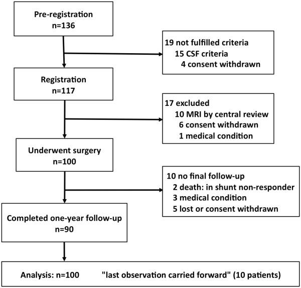
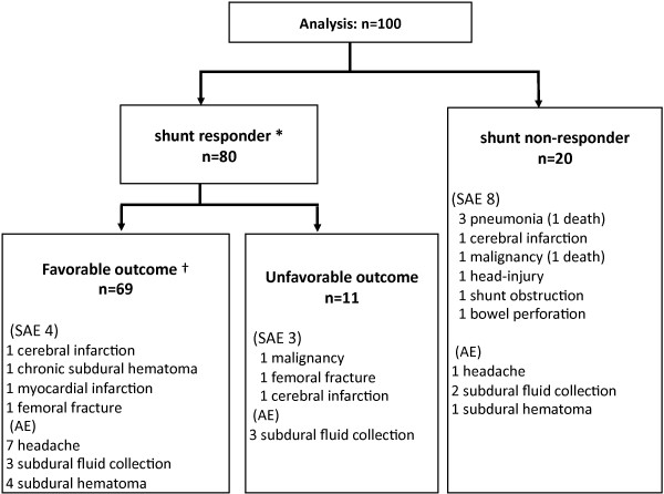
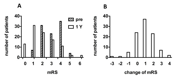
Methods: Twenty-six centers in Japan were involved in this study. Patients aged between 60 and 85 years with one or more of symptoms (gait, cognitive, and urinary problems) and MRI evidence of ventriculomegaly and tight high-convexity and medial subarachnoid spaces received VP shunt using the height/weight-based valve pressure-setting scheme. The primary endpoint was a favorable outcome (improvement of one level or more on the modified Rankin Scale: mRS) at one year after surgery, and the secondary endpoints included improvement of one point or more on the total score of the iNPH grading scale. Shunt responder was defined by more than one level on mRS at any evaluation point in one year.
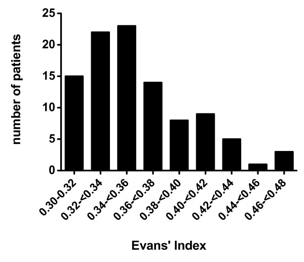
Results: The full analysis set included 100 patients. A favorable outcome was achieved in 69.0% and 80.0% were shunt responders. When measured with the iNPH grading scale, the one-year improvement rate was 77.0%, and response to the surgery at any evaluation point was detected in 89.0%. Serious adverse events were recorded in 15 patients, three of which were events related to surgery or VP shunt. Subdural effusion and orthostatic headache were reported as non-serious shunt-related adverse events, which were well controlled with readjustment of pressure.
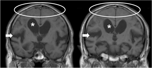
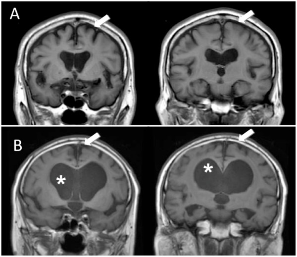
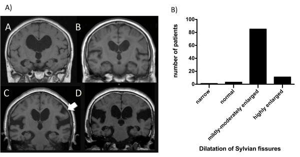
Conclusions: The MRI-based diagnostic scheme is highly useful. Tight high-convexity and medial subarachnoid spaces, and enlarged Sylvian fissures with ventriculomegaly, defined as disproportionately enlarged subarachnoid-space hydrocephalus (DESH), are worthwhile for the diagnosis of iNPH. This study is registered with ClinicalTrials.gov, number NCT00221091.
Figures
References
-
- Guideline Committee, Japanese Society of Normal Pressure Hydrocephalus. Clinical guidelines for idiopathic normal pressure hydrocephalus. Osaka: Medical Review; 2004.
-
- Ishikawa M, Hashimoto M, Kuwana N, Mori E, Miyake H, Wachi A, Takeuchi T, Kazui H, Koyama H. Guidelines for management of idiopathic normal pressure hydrocephalus: Guidelines from the Guidelines Committee of Idioparthic Normal Pressure Hydrocephalus, the Japanese Society of Normal Pressure Hydrocephalus. Neurol Med Chir (Tokyo) 2008;48(Suppl):S1–S23. doi: 10.2176/nmc.48.S1. - DOI - PubMed
Associated data
LinkOut - more resources
Full Text Sources
Medical
