

Steroid hormones content and proteomic analysis of canine follicular fluid during the preovulatory period
- PMID: 21040564
- PMCID: PMC2990747
- DOI: 10.1186/1477-7827-8-132
Steroid hormones content and proteomic analysis of canine follicular fluid during the preovulatory period
Abstract
Background: Follicular fluid contains substances involved in follicle activity, cell differentiation and oocyte maturation. Studies of its components may contribute to better understanding of the mechanisms underlying follicular development and oocyte quality. The canine species is characterized by several ovarian activity features that are not extensively described such as preovulatory luteinization, oocyte ovulated at the GV stage (prophase 1) and poly-oocytic follicles. In this study, we examined the hypothesis that the preovulatory LH surge is associated with changes in steroid and protein content of canine follicular fluid prior to ovulation.
Methods: Follicular fluid samples were collected from canine ovaries during the preovulatory phase, before (pre-LH; n = 16 bitches) and after (post-LH; n = 16) the LH surge. Blood was simultaneously collected. Steroids were assayed by radioimmunoassay and proteomic analyses were carried out by 2D-PAGE and mass spectrometry.
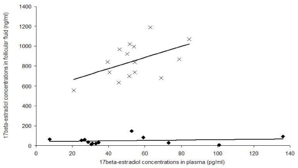
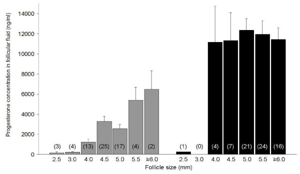
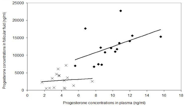
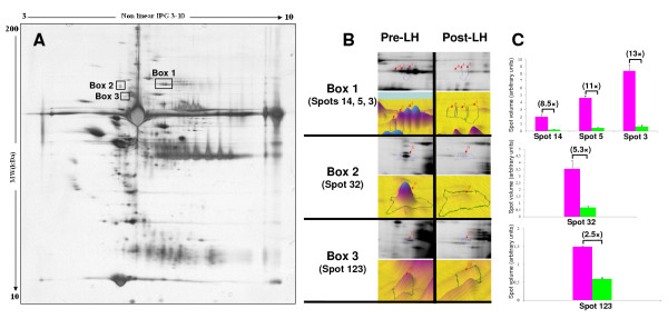
Results: The concentrations of 17beta-estradiol and progesterone at the pre-LH stage were 737.2 +/- 43.5 ng/ml and 2630.1 +/- 287.2 ng/ml in follicular fluid vs. 53 +/- 4.1 pg/ml and 3.9 +/- 0.3 ng/ml in plasma, respectively. At that stage, significant positive correlations between follicular size and intra-follicular steroid concentrations were recorded. After the LH peak, the intrafollicular concentration of 17beta-estradiol decreased significantly (48.3 +/- 4.4 ng/ml; p < 0.001), whereas that of progesterone increased (11690.2 +/- 693.6 ng/ml; p < 0.001). Plasmatic concentration of 17beta-estradiol was not modified (49 +/- 9.6 pg/ml) after the LH peak, but that of progesterone significantly increased (9.8 +/- 0.63 ng/ml).Proteomic analysis of canine follicular fluid identified 38 protein spots, corresponding to 21 proteins, some of which are known to play roles in the ovarian physiology. The comparison of 2D-PAGE patterns of follicular fluids from the pre- and post-LH stages demonstrated 3 differentially stained single spot or groups of spots. One of them was identified as complement factor B. A comparison of follicular fluid and plasma protein patterns demonstrated a group of 4 spots that were more concentrated in plasma than in follicular fluid, and a single spot specific to follicular fluid. These proteins were identified as gelsolin and clusterin, respectively.
Conclusion: Our results provide the first demonstration of size-related changes in the steroid concentrations in canine follicular fluid associated with the LH surge. 2D protein mapping allowed identification of several proteins that may play a role in follicle physiology and ovarian activity at the preovulatory stage. This may help in the future to explain and to better understand the species specificities that are described in dogs.
Figures


Similar articles
-
Estradiol, progesterone, testosterone profiles in human follicular fluid and cultured granulosa cells from luteinized pre-ovulatory follicles.Reprod Biol Endocrinol. 2010 Oct 11;8:117. doi: 10.1186/1477-7827-8-117. Reprod Biol Endocrinol. 2010. PMID: 20937107 Free PMC article. Clinical Trial.
-
Frequency of luteinizing hormone pulses in cattle influences duration of persistence of dominant ovarian follicles, follicular fluid concentrations of steroids, and activity of insulin-like growth factor binding proteins.Anim Reprod Sci. 2003 Jul 15;77(3-4):187-211. doi: 10.1016/s0378-4320(03)00038-1. Anim Reprod Sci. 2003. PMID: 12695054 Clinical Trial.
-
Levels of follicle-stimulating hormone, luteinizing hormone, oestradiol-17 beta and progesterone, and follicular growth in the pseudopregnant rat.J Endocrinol. 1975 Jan;64(1):37-47. doi: 10.1677/joe.0.0640037. J Endocrinol. 1975. PMID: 1167896
-
Preovulatory follicle contributions to oocyte competence in cattle: importance of the ever-evolving intrafollicular environment leading up to the luteinizing hormone surge.J Anim Sci. 2022 Jul 1;100(7):skac153. doi: 10.1093/jas/skac153. J Anim Sci. 2022. PMID: 35772757 Free PMC article. Review.
-
Ovarian follicular and luteal physiology.Int Rev Physiol. 1980;22:117-201. Int Rev Physiol. 1980. PMID: 6248477 Review.
Cited by
-
Seasonal variation in equine follicular fluid proteome.Reprod Biol Endocrinol. 2019 Mar 6;17(1):29. doi: 10.1186/s12958-019-0473-z. Reprod Biol Endocrinol. 2019. PMID: 30841911 Free PMC article.
-
A proteomic analysis of human follicular fluid: comparison between younger and older women with normal FSH levels.Int J Mol Sci. 2014 Sep 29;15(10):17518-40. doi: 10.3390/ijms151017518. Int J Mol Sci. 2014. PMID: 25268621 Free PMC article.
-
Meiotic Development of Canine Oocytes from Poly-Ovular and Mono-Ovular Follicles after In Vitro Maturation.Animals (Basel). 2023 Feb 13;13(4):648. doi: 10.3390/ani13040648. Animals (Basel). 2023. PMID: 36830434 Free PMC article.
-
Proteomic analysis of mare follicular fluid during late follicle development.Proteome Sci. 2011 Sep 17;9:54. doi: 10.1186/1477-5956-9-54. Proteome Sci. 2011. PMID: 21923925 Free PMC article.
-
Investigating media that support red wolf (Canis rufus) sperm viability and capacitation in vitro.Reprod Fertil. 2020 Dec 28;1(1):83-92. doi: 10.1530/RAF-20-0042. eCollection 2020 Jul. Reprod Fertil. 2020. PMID: 35128425 Free PMC article.
References
-
- Concannon PW, McCann JP, Temple M. Biology and endocrinology of ovulation, pregnancy and parturition in the dog. J Reprod Fertil Suppl. 1989;39:3–25. - PubMed
Publication types
MeSH terms
Substances
LinkOut - more resources
Full Text Sources
Research Materials
