Review
. 2010 Nov 1:12:e35.
doi: 10.1017/S1462399410001675.

Novel insights into molecular mechanisms of abruption-induced preterm birth

Affiliations
Review

Novel insights into molecular mechanisms of abruption-induced preterm birth

Catalin S Buhimschi et al. Expert Rev Mol Med. .

Abstract

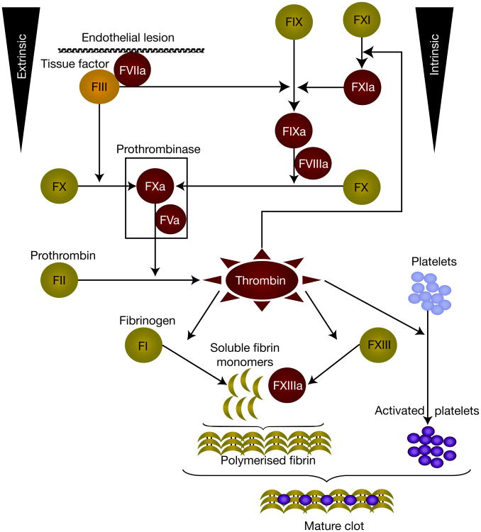
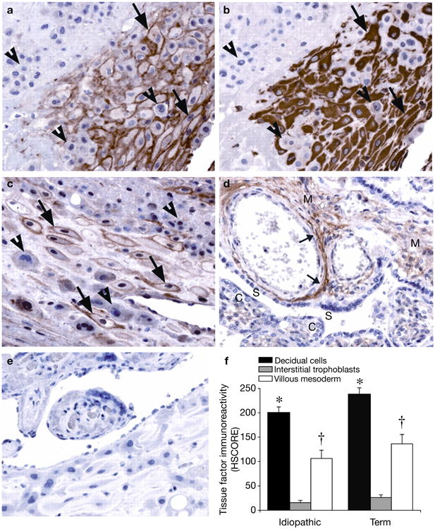
Preterm birth (PTB) complicates more than 12% of all deliveries. Despite significant research, the aetiology of most cases of PTB remains elusive. Two major antecedents of PTB, intra-amniotic infection and decidual haemorrhage (abruption), can exhibit dissimilar demographic and genetic predispositions, despite sharing common molecular and cellular pathways. The use of high-throughput, high-dimensional technologies reveals substantial crosstalk between the coagulation and inflammation pathways. Tissue factor, thrombin and cytokines are key mediators of this crosstalk. Abruptions are associated with excess thrombin generated from decidual-cell-expressed tissue factor. Although thrombin is a primary mediator of the coagulation cascade, it can also promote inflammation-associated PTB by enhancing expression of matrix metalloproteinase and neutrophil-chemoattracting and -activating chemokines. Here, we provide novel insights into the molecular mechanisms and pathways leading to PTB in the setting of placental abruption.


Figures

Figure 1
Figure 1. Extrinsic and intrinsic pathways of the coagulation cascade
Tissue factor (TF) is recognised as the main initiator of the coagulation cascade. Once formed, the TF–FVIIa complex initiates clotting by activating both FX and FIX thus, TF carries a unique ability to act through both intrinsic and extrinsic pathways of coagulation. Activated FIX binds to its cofactor VIIIa, to activate FX. Thus, the FVIIIa–FIXa complex of the intrinsic pathway provides an alternative route to generate FXa, which participates in the prothrombinase complex (FVa–FXa). The latter complex activates prothrombin to thrombin, which has a central role in the coagulation protease cascade. Thrombin, in turn, activates FXI, which is an alternative way of generating FIXa. Thrombin also activates FXIII, which stabilised fibrin polymers, but thrombin's most crucial functions are to cleave fibrinogen to fibrin and stimulate platelets by the cleavage of protease-activated receptors. The end result is haemostasis and thrombosis. Abbreviations: FI, factor I; FII, factor II; FIII, factor III; FV, factor V; FVII, factor VII; FIX, factor IX; FX, factor X.
Figure 2
Figure 2. Immunohistochemical analysis of tissue factor expression at the decidual–placental interface
Serial sections of decidual basalis specimens immunostained for tissue factor (TF) and vimentin, as shown in an idiopathic preterm specimen: (a) TF and (b) vimentin. Decidual cells (arrows), identified by positive vimentin staining, exhibit strong perimembranous TF staining. TF staining is absent in the interstitial trophoblast (arrowheads). Similar results are seen in term specimens (c). Placental villi show moderate TF staining in the mesoderm (M), in which the staining is primarily localised to perivascular adventitia (small arrows), as shown in a preterm specimen (d). The syncytiotropoblast (S) and cytotrophoblast (C) show no TF staining. Similar results are seen in term specimens (not shown). Negative control immunostaining using a nonspecific isotype-matched antibody revealed no positive signals (e). TF staining intensity (f) [bars indicate histological score (HSCORE) mean ± s.e.m.] is highest in decidual cells (*P<0.05, versus interstitial trophoblast and the villous mesoderm of the same specimen group), with moderate staining in the villous mesoderm (P<0.05, versus interstitial trophoblast of the same specimen group). There are no significant differences in HSCORE values between specimen groups. Published with permission from Ref. (© 2009, The Endocrine Society).
Figure 3
Figure 3. Representative proteomic profiles characteristic for intra-amniotic inflammation and bleeding
Three hypervariable areas of representative surface-enhanced laser desorption ionisation time-of-flight tracings of amniotic fluid from women admitted for preterm birth are shown: (a) 3–4 kDa, (b) 10–12.5 kDa and (c) 14–17 kDa. These areas contain proteomic patterns characteristic of intra-amniotic inflammation (MR score composed of four protein biomarkers – P1, P2, P3 and P4) (Ref. 79), intra-amniotic bleeding (Hb chains) or both (bottom tracings). The x-axis of the tracings represents the molecular mass in daltons; the y-axis represents the normalised peak intensity. R denotes a reference protein peak present in all fluid samples, which corresponds to a fragment of beta-2 microglobulin (originally published in Ref. ; reproduction permitted under Creative Commons Attribution License). Abbreviations: Hb, haemoglobin; MR, mass-restricted; P1, neutrophil defensin-2; P2, neutrophil defensin-1; P3, calgranulin C; and P4, calgranulin A.
Figure 4
Figure 4. Binary theory of decidual bleeding and inflammation in pathogenesis of placental abruption
A reduced or intermittent uteroplacental blood flow causes focal decidual hypoxia and free radicals and ROS (through reperfusion injury). Hypoxia induces the expression of decidual VEGF. This angiogenic factor acts directly on decidual endothelial cells to enhance permeability and degrade the vascular wall through MMP-2 generation. This leads to haemorrhage and aberrant endothelial expression of TF to generate additional thrombin that further induces TF expression and uteroplacental thromboses, which exacerbate reduced blood flow. Free radicals and ROS induce endothelial cell injury, which allows perivascular leakage of coagulation factors, including factor VIIa, which then comes in contact with TF, activating the extrinsic coagulation pathway. The resulting thrombin further induces TF expression as well as expression of inflammatory cytokines, leading to inflammation in the absence of a microbial attack. Abbreviations: MMP, matrix metalloproteinase; ROS, reactive oxygen species; sFlt-1: tyrosine kinase-1; TF, tissue factor; VEGF, vascular endothelium growth factor.

References

    1. Stoll BJ, et al. National Institute of Child Health and Human Development Neonatal Research Network. Neurodevelopmental and growth impairment among extremely low-birth-weight infants with neonatal infection. Journal of the American Medical Association. 2004;292:2357–2365. - PubMed
    1. Zerhouni EA. US biomedical research: basic, translational, and clinical sciences. Journal of the American Medical Association. 2005;294:1352–1358. - PubMed
    1. Fagnan LJ. Linking practice-based research networks and clinical and translational science awards: new opportunities for community engagement by academic health centers. Academic Medicine. 2010;85:476–483. - PMC - PubMed
    1. McLean M, et al. A placental clock controlling the length of human pregnancy. Nature Medicine. 1995;1:460–463. - PubMed
    1. Zenclussen AC, et al. Immunology of pregnancy: cellular mechanisms allowing fetal survival within the maternal uterus. Expert Reviews in Molecular Medicine. 2007;9:1–14. - PubMed

Publication types